Image quality of a proton radiography system using 2D lateral projections for image guidance
Mikael Simard,
United Kingdom
OC-0774
Abstract
Image quality of a proton radiography system using 2D lateral projections for image guidance
Authors: Mikael Simard1, Daniel Robertson2, Ryan Fullarton1, Allison Toltz3, Colin Baker3, Sam Beddar4, Charles-Antoine Collins-Fekete1
1University College London, Medical Physics & Biomedical Engineering, London, United Kingdom; 2Mayo Clinic Arizona, Division of Medical Physics, Department of Radiation Oncology, Phoenix, USA; 3University College London Hospital, NHS Foundation Trust, Radiotherapy Physics, London, United Kingdom; 4The University of Texas MD Anderson Cancer Center, Radiation Physics, Houston, USA
Show Affiliations
Hide Affiliations
Purpose or Objective
Image guidance is a necessary step to help proton therapy reach its full potential for the treatment of cancers such as non-small cell lung cancer. Using the treatment source to capture projection proton radiographs (pRads) is an attractive imaging solution, as it is registered with the treatment beam and limits water equivalent thickness (WET) errors. Proton imaging is typically achieved using single-event imaging, which is currently too slow for practical image guidance and generally not compatible with clinical beam settings. An alternative, integrated mode proton imaging, consists of acquiring signals from individual pencil beams (PB) using clinical beam parameters. The objective of this work is to report imaging quality of a low-cost and fast imaging device based on a volumetric scintillator and CCD cameras for integrated mode pRads. Image reconstruction is achieved by fully exploiting captured 2D lateral projections of the dose deposition in the scintillator.
Material and Methods
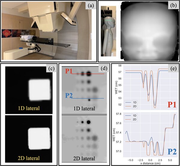
Proton radiographs were acquired at Mayo Clinic Arizona using a plastic volumetric scintillator and a single CCD camera capturing a projected 2D lateral view of 3D energy deposition in the scintillator (Fig 1a). The device covers a 13x13 cm2 field of view. Image reconstruction is done using a novel reconstruction framework which identifies multiple candidate Bragg peaks in lateral views and performs a weighted reprojection of the WET according to Fermi-Eyges theory. Data was acquired using clinical settings at 135 MeV, and PB spacings of 2, 3, 4 and 5 mm. Results are presented for a 3 mm spacing which is a compromise between imaging time and quality. The following phantoms were scanned: Las Vegas (contrast), slanted edge (resolution), 9 tissue-substitute Gammex inserts (quantitative WET accuracy), and a paediatric head phantom. The reconstruction using projected 2D lateral views is compared with a conventional integrated mode system (1D Bragg curve).
Results
Clinical image quality of our reconstruction for the paediatric head phantom is shown in fig 1b. Compared to conventional integrated mode approach, our method yields a 30% relative increase in resolution (fig 1c. defined as 10% of the modulation transfer function) and a 60% relative increase of contrast (Fig 1d-e). Fig 2 illustrates an excellent WET accuracy (mean absolute error of 0.4 mm over all 9 inserts). Sub-second imaging can be achieved with a 6 mm beam sampling.
 à
à
Figure 1 - image quality (resolution, contrast) for the integrated mode proton radiography system
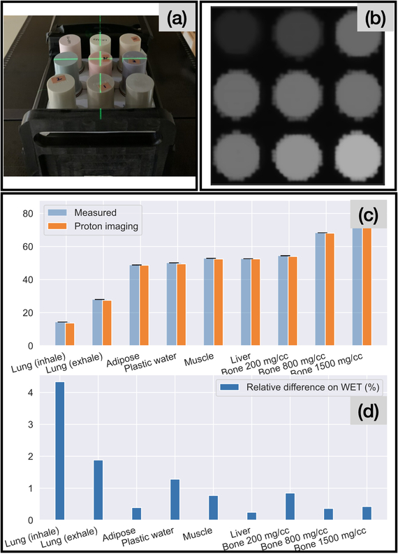
Figure 2 - WET accuracy for the integrated mode proton radiography system
Conclusion
This work illustrates that high quality/clinically ready proton radiographs can be obtained with clinical beam settings using a fast, low-cost scintillation-based system, and shows benefits of 2D Bragg curve data as opposed to a range telescope for integrated mode pRads. Future work includes rapid fluoroscopic imaging for real-time treatment adaptation, and potential use for proton therapy QA.