PSD (plastic scintillation detector) in vivo dosimetry in post-operative endometrial brachytherapy
PO-1802
Abstract
PSD (plastic scintillation detector) in vivo dosimetry in post-operative endometrial brachytherapy
Authors: Antonio Herreros1, Joana Melo2, Luis Moutinho2
1Hospital Clínic Barcelona, Radiation Oncology. Medical Physics Section, Barcelona, Spain; 2NU-RISE, Lda, Aveiro, Portugal
Show Affiliations
Hide Affiliations
Purpose or Objective
It has been estimated that only around 20-30% of European hospitals with brachytherapy perform in vivo dosimetry. However, in brachytherapy there are still many manual procedures that could derive in adverse events, and which could be detected with in vivo dosimetry. PSD (Plastic scintillator detectors) have interesting properties such as good spatial resolution, real-time reading, linearity, water equivalence, low dependence on temperature, watertight, low angular polar and azimuthal dependence, low dependence on energy, radiation resistance, short-term repeatability, low dependence on dose rate and sizes smaller than 1mm. The purpose of this study was to evaluate the performance of a PSD in post-operative endometrial brachytherapy in terms of dwell time accuracy.
Material and Methods
The PRODOSE in vivo dosimetry system (NU-RISE, Portugal) uses an organic scintillator BCF-12 (Saint Gobain Crystals, France) with a length of 2 mm attached to a PMMA optic fiber with a diameter of 0.5mm. The detection technology consists in a silicon photomultiplier matrix (SiPMs). The system works with an automatic temperature correction.
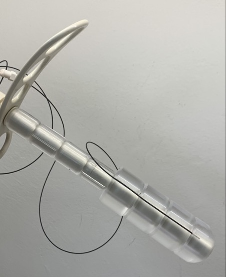
In a previous work, measurements were performed in a bladder catheter, but uncertainties in the detector positioning in this setup has led us to develop a drilled vaginal cylinder applicator to insert the PSD probe near to the surface of the applicator and have a more stable geometry. In Figure 1, a view of the drilled applicator with the PSD probe inside can be visualized. The measurements were carried out in 19 sessions of 17 patients treated with an afterloader microSelectron v3 Digital (Elekta, The Netherlands). In four patients the described drilled vaginal cylinder applicator was used to irradiate the vaginal cuff. The treatment plans were calculated using Oncentra Brachy TPS v.4.5.3 (Elekta, The Netherlands) after importing the 0.6 mm spaced axial images from a Siemens Somatom go.Open Pro CT (Siemens Healthineers, Germany).
Although the signal can be visualized on real time, the raw measurements were processed with a MATLAB R2014a (The MathWorks, Inc., Massachusetts) script to obtain the times of each dwell position and compare with the TPS calculated values.
Results
The average deviation between the total treatment plan dwell times and the measured ones is lower than 0.1% (0.08%), with an maximum deviation of 0.28%, a median of 0.06% and a standard deviation of 0.11%.
Conclusion
In vivo PSD determines accurately the dwell times, and it is a quality assurance procedure that allows verification of dose delivery in each fraction. In view of the good results obtained in time measurements with the PSD detector, a robust calibration procedure is being designed to calibrate the PSD detector in terms of absorbed dose.