A DECT framework to measure the effect of radiation dose on lung function after radiotherapy
Mikael Simard,
United Kingdom
PO-1748
Abstract
A DECT framework to measure the effect of radiation dose on lung function after radiotherapy
Authors: Mikaël Simard1,2, Andréanne Lapointe3, Houda Bahig3, Jean-François Carrier1,2,3, Shen Zhang3, Marie-Pierre Campeau3, Édith Filion3, David Roberge3, Hugo Bouchard1,2,3, Stéphane Bedwani3,2
1Université de Montréal, Physique, Montréal, Canada; 2Centre hospitalier de l’Université de Montréal, Centre de recherche, Montréal, Canada; 3Centre hospitalier de l’Université de Montréal, Radio-oncologie, Montréal, Canada
Show Affiliations
Hide Affiliations
Purpose or Objective
To propose a contrast-enhanced dual-energy CT (DECT) framework to study dose effect on lung function after radiotherapy and validate the usefulness of the framework with a case study of four patients.
Material and Methods
Four lung cancer patients received a DECT scan (Siemens SOMATOM Definition Flash) with intravenous iodine injection before the radiotherapy treatment (τ=0), and at τ=6 and τ=12 months post-treatment. Post-treatment DECT scans were registered on the pre-treatment scans using the Advanced Normalization Tools (ANTs) software to allow correspondence with the dose map. Iodine maps were estimated with a two-material decomposition using lung and iodine as basis materials, and a stoichiometric calibration procedure.
As iodine concentration may vary importantly between patients and times τ, it is not a purely quantitative metric. A different metric, the normalized function (NF), is reported. The NF is defined as the ratio of mean iodine concentration in a given region of interest normalized by the mean iodine concentration in a reference region. The reference region is defined as a well perfused volume that received low dose (under 5 Gy). Furthermore, the functional response (FR) is defined as the relative variation of the NF between pre-treatment and post-treatment scans.
Results
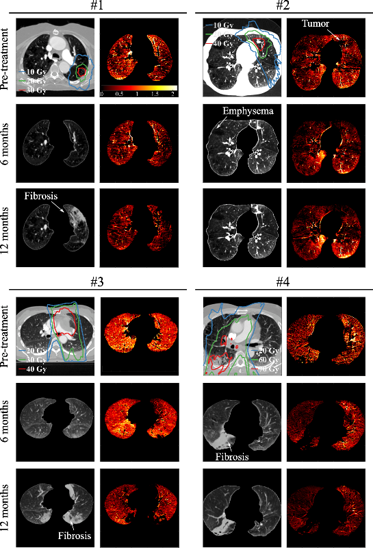
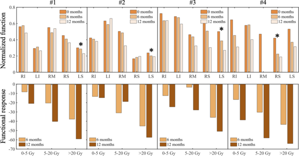
Figure 1 shows examples of fully registered CT images along with the corresponding functional data (NF images) for each of the four patients and τ=0, 6 and 12 months. The apparition of fibrosis (patients 1 and 3) or the general loss of function after treatment (patients 3 and 4) is recognizable. Figure 2 shows the metrics NF and FR for selected regions of interest. On the top row, the NF is shown for each of the five lung lobes (right and left inferior RI and LI, right middle RM, right and left superior RS and LS). Tumor location for all patients correlate with poor NF at τ=12 months compared to other lobes, suggesting that the loss of function is important close to the tumor.
The bottom row of figure 2 shows the FR for three dose ranges (<5 Gy, 5-20 Gy, >20 Gy) for each patient. It is shown that lung regions which received higher dose are generally associated with a more important loss of function over time. The metric thus allows comparing the relative response to radiation between patients.

Fig 1 - examples of fully registered CT images (gray colormap) and corresponding functional data (NF heat maps) for all patients and times τ.

Fig 2 - for all patients, (top) NF for each lung lobe, (bottom) FR for selected dose ranges. The location of the tumor is identified with an asterisk.
Conclusion
The proposed methodology is suitable to follow the evolution of lung function after treatment. The NF and FR can be used as metrics to identify regions with low functionality and poor response to radiation. Further work will apply this methodology to a large cohort study of >50 patients and up to 24 months follow-up, with the long-term objective to limit and predict possible complications following lung radiotherapy.