An analytic method for inhomogeneity correction of Gafchromic EBT3 films
PO-1630
Abstract
An analytic method for inhomogeneity correction of Gafchromic EBT3 films
Authors: Marco Parisotto1, Vittoria Emanuela Morabito1, Stefano Ferretti1, Luca Reversi1, Francesco Cesarini1, Marco Valenti1
1Ospedali Riuniti di Ancona, Medical Physics, Ancona, Italy
Show Affiliations
Hide Affiliations
Purpose or Objective
An analytic method for inhomogeneity correction of Gafchromic EBT3 films was developed by the authors and evaluated. Correction factors to single-channel dosimetry was provided to account for Later Effect Artifact (LRA) and film thickness variation.
Material and Methods
Single-channel dosimetry correction factors
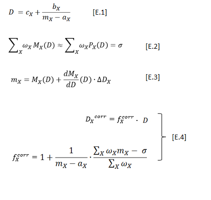
Single-channel (SC) dosimetry was expressed by [E.1], where m was the scanner readings of uniformely exposed Grafchromic EBT3 films to dose D, the subscript X represented a channel color among RBG and a,b,c some fitting parameters. Inversion of [E.1] represented the calibration curves M(D) of the films [1]. We calculated the coefficients of three second-order polynomials P(D) which best approximated the calibration curves in the dose-range 0-3 Gy. In such a way, some weights w which satisfied [E.2] did exist, where sigma was an arbitrarily constant value. Considering [E.3] in the first order development of M around D, we found that dose could be expressed by [E.1] multiplied by the correction factor in [E.4]. Expression [E.4] then represented the "corrected" single-channel (CSC) dosimetry.

Film exposures
Three 25x25 cm2 Grafchromic EBT3 films were irradiated (Varian 21EX, 6MV) in a RW3 phantom with an open beam of 20x20 cm2 size at film plane. The calculated dose (Varian Eclipse with AAA) at central axis were 72 cGy, 144 cGy and 287 cGy, respectively, The films were digitalized 24-hours after exposure using a multi-channel flatbed scanner Epson Expression 10000XL. The films belonged to a lot previously calibrated using [E.1]. Image to dose conversion was performed applying the calibration curves for single-channel (SC) dosimetry. Correction factors were then calculated using the proposed method. The effectiveness of the "corrected single-channel" (CSC) dosimetry in accounting for LRA effect was evaluated against the central axis dose profiles extracted perpendicularly to scan direction.
Results
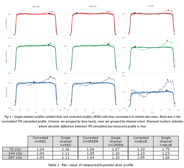
The proposed method showed an evident mitigation of LRA for the central axis dose profiles extracted perpendicularly to scan direction (fig. 1). In table 1 the maximum values of measured/expected dose ratio among different channels were reported.

Conclusion
The proposed method provided a more reliable dosimetry respect to single-channel dosimetry,