PTV changes analysis throughout treatment in H&N patients
Laura Cardoso Rubio,
Spain
PO-1118
Abstract
PTV changes analysis throughout treatment in H&N patients
Authors: Sofía Pena Vaquero1, Laura Cardoso Rubio2, Angel del Castillo Belmonte1, Laura Gómez heras2, Iban Conles Picos1, Antonio Hurtado Romero1, David Miguel Pérez1, Delfín Alonso Hernández1, Ricardo Torres Cabrera1
1Hospital Clínico Universitario, Medical Physics, Valladolid, Spain; 2Hospital Clínico Universitario, Radiation Oncology, Valladolid, Spain
Show Affiliations
Hide Affiliations
Purpose or Objective
Image-guided
radiotherapy (IGRT) is commonly used to minimize the effect of positioning and
to control tumor response. It is well known that interfraction variations can significantly
influence the dose received by the lesion or organs at risk. These differences
are sensitive in treatments such as in head and neck (H&N) region.
An analysis
of planning target volumes (PTV) size changes in H&N treatments through
image deformation tools is presented.
Material and Methods
Seven patients
with head and neck pathology treated with VMAT and IGRT by CBCT were retrospectively
selected. The total number of PTV was 18, which were distributed among the
seven patients with two or three prescribed dose levels ranging from 70 to 54
Gy.
To evaluate
PTV volume changes, a synthetic CT (sCT) is generated from the CBCT image for
each session. For this purpose, a deformable fusion algorithm implemented in VelocityTM v4.1 is
employed to modify the planning CT and the contours using the online match
performed between daily CBCT and the planning CT as a basis.
Results
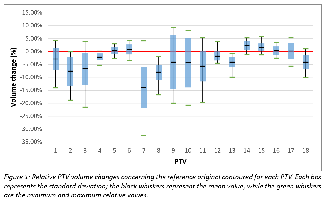
The size
variation across the treatment by the PTVs with respect to the originally
contoured ones implies that the volume is smaller, on average. This average
shows a decrease of over 3%, with a standard deviation of 4% (Figure 1).

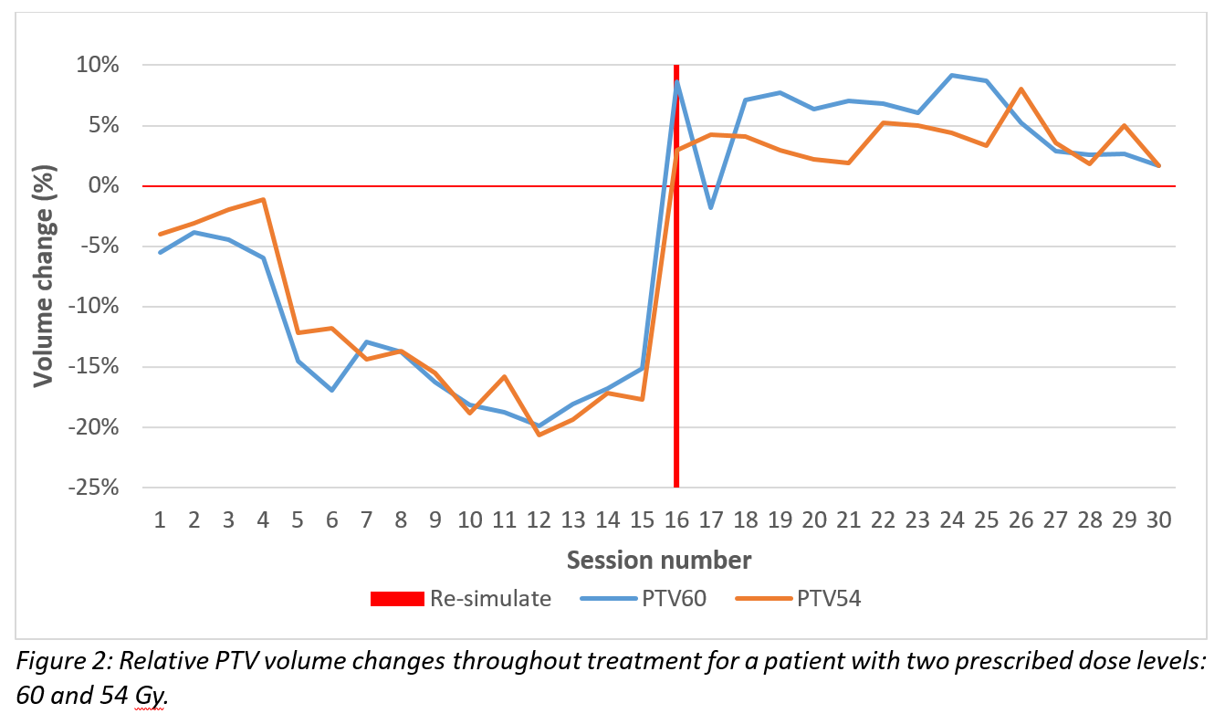
To illustrate
how changes affect during a course, as an example, figure 2 shows daily changes
on a patient.

Conclusion
A
procedure for monitoring anatomical changes during treatment has been shown,
warning about meaningful volume differences and helping us to make clinical
decisions on treatment execution.