Dosimetric consistency between MR-only and deformed CT for MR-guided online adaptive radiotherapy
Grichar Valdes Santurio,
Denmark
OC-0775
Abstract
Dosimetric consistency between MR-only and deformed CT for MR-guided online adaptive radiotherapy
Authors: Grichar Valdes Santurio1, Jens Edmund1, Katri Ilona Nousiainen2
1Herlev Hospital, Radiotherapy, Herlev, Denmark; 2Helsinki University, Helsinki University Hospital, Helsinki, Finland
Show Affiliations
Hide Affiliations
Purpose or Objective
MR-guided online adaptive radiotherapy (MRgOART) has
become more frequent in radiotherapy clinics with the inclusion of MR-linacs
(MRL). In the workflows for adaptive fractions, an MR scan is first acquired
using a dedicated MRL sequence, the structure set re-contoured, and the plan
re-optimized and re-calculated. For plan re-calculation, an electron densities
(ED) map is assigned from either the deformed or rigid CT simulated before
treatment. The ED map extracted from the deformed CT (dCT) is not necessarily
consistent with the MR image and thus subject to QA prior the treatment delivery.
To assess the dosimetric impact of incorrect ED alignment with the MRI, bulk EDs
were manually assigned to the MR images and the plan was re-calculated using
those 2 conditions. The bulk ED assignment is typically called MR-only
Material and Methods
The 0.35T MRL from ViewRay was used for calculating
the plans and acquiring MR images. In this system, a Monte Carlo based
treatment planning system uses the ED relative to water (RED) for material
assignment and calculating dose-to-medium. 2 bulk ED were manually assigned:
“water-like” for all the structures inside the patient (RED=1), and “lung-like”
for the lungs (RED=0.12). 15 patients were included with lesions mainly located
in the upper-abdominal/thoracic regions such as pancreas and liver. 1
online-adaptive fraction per patient was selected as a representation of the
full treatment. The 15 patients exhibit 32 PTV targets and 216 organs at risk
(OARs) in total. Several points of the dose volume histogram (DVH) were compared;
the 98%, 50% and 2% (near, median and max coverage) for the targets, and the 2%
and mean dose (serial and parallel type indicators) for the OARs. The
discrepancies were normalized relative to the prescribed dose
Results
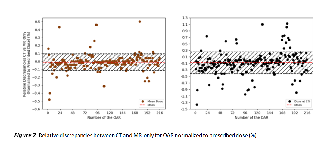
Figures 1 and 2 show the relative discrepancies between the doses for the MR-only
and CT approach. The dash red lines and bands indicate mean and ±1SD of the data.
For all target DVH points in figure 1, all discrepancies are <2% with a
systematic shift in mean value of -0.16% For the 2 OARs DVH points, the 2 the
highest discrepancy is <1.4% and mean value <-0.08%. In general, the deviations
for the OARs show less spread as compared to the targets which suggests less
dosimetric uncertainty (more robustness for ED assignment) of the OARs than for
the targets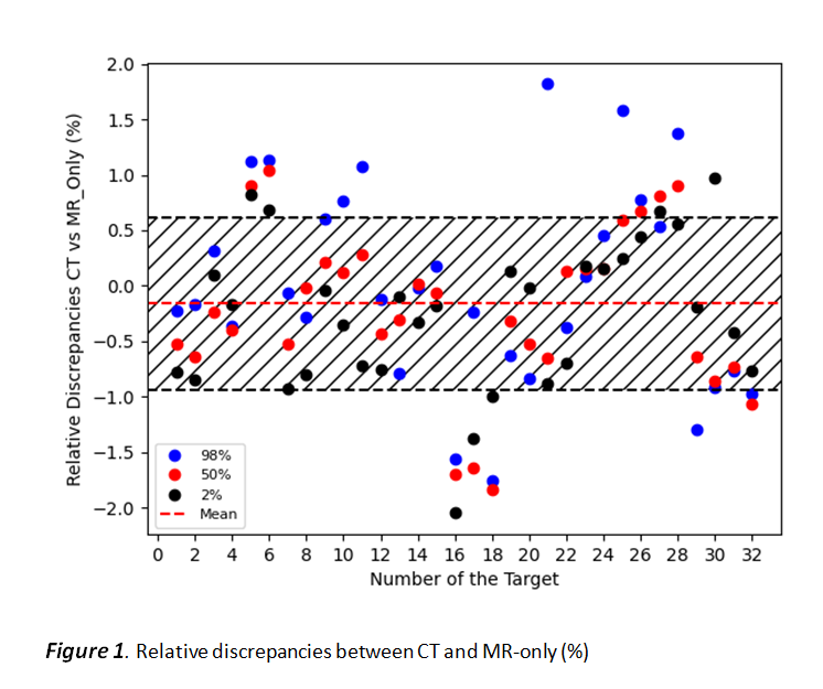
Conclusion
This study evaluates the possibility of using MR-only for MRgOART in the
upper/abdominal-thoracic region. With MR-only using assigned ED of water and
lung, the discrepancies with the CT is less than 2% and 1.4% for the targets and
OARs, respectively. Therefore, variations of less than 2% are expected when the
CT-based ED map does not follow the MR image in adaptive radiotherapy workflows
for the upper abdominal/thoracic region. We believe that the way forward is to
assign an RED for all the structures based on e.g. the ICRU 46 report or synthetic
CT generation directly from the MRI. This will allow for a more thorough
comparison between ED methods