Correlation between External and Internal Tumour Motion in Lung Stereotactic Ablative Radiotherapy
ASMAA ALI,
United Kingdom
PD-0230
Abstract
Correlation between External and Internal Tumour Motion in Lung Stereotactic Ablative Radiotherapy
Authors: ASMAA ALI1, Jason Greenwood1, Louise Belshaw2, Glenn Whitten2, Denise Irvine2, AR. Hounsell2,3, CK. McGarry3,2
1Queen’s University Belfast, School of Mathematics and Physics, Belfast, United Kingdom; 2Northern Ireland Cancer Centre, Belfast Health and Social Care Trust, Radiotherapy Physics, Belfast, United Kingdom; 3Queen’s University Belfast, Patrick G Johnston Centre for Cancer Research, Belfast, United Kingdom
Show Affiliations
Hide Affiliations
Purpose or Objective
An important challenge when delivering radiotherapy to lung cancer patients is the breathing-induced tumour motion. This issue is crucial when improved geometrical accuracy is required, as in stereotactic ablative radiotherapy (SABR). This study aims to investigate the correlation between the motion from the external surrogate and internal tumour motion.
Material and Methods
A dataset of 260 4DCT scans of lung SABR patients was analysed. The 4DCT images were generated using a GE Optima 580 and a Discovery 590 RT (GE Healthcare, Waukesha, WI), each coupled with the Varian real-time position management system RPM (Varian Medical Systems, Palo Alto, CA). The images were acquired using the cine 4DCT protocol (120 KV, 250 mA), with a slice thickness of 2.5mm. The images were binned into ten breathing phases and imported into the Varian Eclipse treatment planning system (v16.01). All tumours were classified based on their geometrialc location into three anatomical lobes.
The recorded internal tumour information included the centroid motion of the target volume between the end inhalation and end exhalation phases (GTV0% and GTV50%) in the superior-inferior (SI), anterior-posterior (AP), and left-right (LR) directions and the 3D motion. For the external assessment, the RPM breathing signals of all patients were analysed via an in-house script. The median amplitude in the AP direction (external) was defined using the breathing signal during the CT acquisition (beam-on). The external motion was correlated with the internal 3D centre of mass motion measured from the 4DCT via Pearson correlations for each lobe. The Kruskal-Wallis test with Bonferroni adjustment was carried out to determine significant differences in the 3D motion, SI, AP, and the LR directions across the three anatomical lobes (significance set at P<0.05).
Results
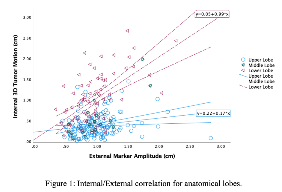
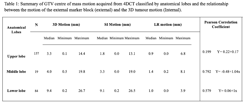
Table 1 shows that there was significantly higher 3D motion in the lower lobe tumours (range: 26.4 mm) compared to the middle (range: 19.3 mm) and upper lobe tumours (range: 14.3 mm). A significant difference was observed in the 3D and SI motion across the anatomical lobes (p<0.001). Between the groups, no significant difference was observed between upper and middle lobe (p>0.6), but there was significant differences between upper and lower lobe (p<0.001), and middle and lower lobe (p<0.02) both for 3D and SI motion. There was no significant difference in the AP and LR motion across anatomical lobes (p=0.41 and p=0.13), respectively. Figure 1 and table 1 show that the external / internal correlation was weak, strong and moderate for the upper, middle and lower lobe tumours, respectively.


Conclusion
The correlation between the external surrogate, which is assumed to be directly related to the diaphragm motion, and the internal tumour motion illustrated a moderate-high correlation for tumours proximal to the diaphragm. The correlation is low among tumours in the upper lobe of the lung.