3D abdominal organ motion correlates strongly with the diaphragm during prolonged breath-holds
Jeffrey Veldman,
The Netherlands
PD-0229
Abstract
3D abdominal organ motion correlates strongly with the diaphragm during prolonged breath-holds
Authors: Jeffrey Veldman1, Zdenko van Kesteren1, Ebony Gunwhy1, Michael Parkes1, Markus Stevens2, Joost van den Aardweg3, Geertjan van Tienhoven4, Arjan Bel5, Irma van Dijk6
1Amsterdam UMC - Location AMC, Department of Radiation Oncology, Amsterdam, The Netherlands; 2Amsterdam UMC - Locations AMC and VUmc, Department of Anaesthesiology, Amsterdam, The Netherlands; 3Amsterdam UMC, Department of Pulmonology, Amsterdam, The Netherlands; 4Amsterdam UMC, Department of Radiation Oncology, Amsterdam, The Netherlands; 5Amsterdam UMC, Department of Radiotherapy, Amsterdam, The Netherlands; 6Amsterdam UMC - Location AMC, Department of Radiotherapy, Amsterdam, The Netherlands
Show Affiliations
Hide Affiliations
Purpose or Objective
Respiratory motion management (RMM) is
recommended for tumours subjected to excursions of more than 5 mm. Prolonged
breath-holds (PBH) of >5 minutes can be achieved by mechanical ventilation
induced hypocapnia after preoxygenation. During these prolonged breath-holds,
the lungs gradually deflate causing the right diaphragm to drift cranially by
approximately 3.0 mm/min. Here, we studied the correlation between the
diaphragm drift and abdominal organ motion using deformable image registration
(DIR) during prolonged breath-holds. Furthermore, we studied the correlation
between the right diaphragm top and abdominal organ motion, to investigate its
applicability as respiratory motion surrogate.
Material and Methods
Seven volunteers performed a total of eleven
PBHs from end-inspiration (four volunteers, two PBHs each; three volunteers,
one PBH each). 3D cine-MRIs of the upper abdomen were acquired with individual 3D
image acquisition duration of 14 s. Seven images (the first, then at 25%, 50%
and 75% of the PBH duration and the last three) were registered using groupwise
DIR resulting in a Deformation Vector Field (DVF) from the first to every other
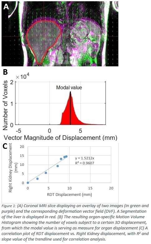
image (example DVF in Figure 1A). Regions-of-interest (ROI) were defined by
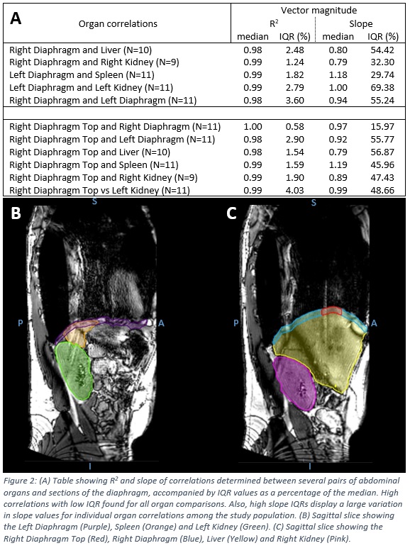
delineating the right and left diaphragm, the right diaphragm top (RDT), liver,
spleen, and both kidneys (Figure 2B and 2C) on the first image. The
distribution of voxel displacements from the first to all other images for each
ROI was computed for the 3-dimensional vector magnitude of the motion resulting
in a Motion Volume Histogram (MVH; Figure 1B). Modal displacements were
extracted from these MVHs, serving as a measure for 3D organ displacement. Correlations
between organ displacements were calculated for each individual PBH for pairs
of organs using a linear regression model (Figure 1C). Median R2 , slope
values and relative inter-quartile ranges (IQR), as percentages of the median
were determined over the eleven PBHs. For each set of organs compared, a
Wilcoxon signed rank test was used to determine if the slope was significantly
different from 1.
Results
Median PBH duration
was 315s (IQR 165s). Abdominal organ motion showed to be highly correlated to
RDT drift within individual PBHs (R2 >0.98) (Figure 2A). Corresponding
IQR values were low (all <5%) displaying small variation over all PBHs. Median
values for the slope were not significantly different from 1, providing no
indication that the diaphragm moves significantly more than the abdominal
organs. High IQRs were found for the slope for numerous organ comparisons
(>45% for 8 of the 11 organ sets) indicating that the relation between RDT
motion and organ motion varies greatly between the different PBHs.
Conclusion
Abdominal organ motion during a single PBH is
highly correlated to the diaphragm and right diaphragm top. The high IQR values
of the slope suggest that the relation between RDT motion and abdominal organ
motion varies between subjects and within subjects.