reconstructing the dosimetric impact of intra-fractional prostate motion in MR-guided radiotherapy
PD-0227
Abstract
reconstructing the dosimetric impact of intra-fractional prostate motion in MR-guided radiotherapy
Authors: Yuqing Xiong1, Moritz Rabe2, Lukas Nierer2, Stefanie Corradini2, Claus Belka2,3, Marco Riboldi4, Guillaume Landry2, Christopher Kurz2
1Univerisity Hospital LMU, Department of Radiation Oncology, Munich, Germany; 2University Hospital LMU, Department of Radiation Oncology, Munich, Germany; 3German Cancer Consortium (DKTK), Partner Site Munich, Munich, Germany; 4LMU Munich, Department of Medical Physics, Munich, Germany
Show Affiliations
Hide Affiliations
Purpose or Objective
This study aimed at evaluating the
intra-fractional prostate motion captured during MR-guided radiotherapy of
prostate cancer and analyzing its impact on the delivered dose over all
treatment fractions.
Material and Methods
Sagittal 2D cine MRI videos were acquired
at 4 Hz during each fraction of MR-guided online adaptive radiotherapy for 10
prostate cancer patients at a 0.35 T MR-linac (ViewRay-MRIdian). The CTV
(prostate) was chosen as the tracking contour and the gating window was defined
as the CTV with an isotropic expansion in the [3, 5] mm range. During
treatment, the target was continuously tracked by the vendor’s optical flow
algorithm. Using the videos and in-house software, the centroid coordinates of
the target were calculated relative to the static gating window in
anterior-posterior (AP) and superior-inferior (SI) direction. Using the static dose
cloud approximation, the planned fraction dose was shifted according to the
extracted motion during the beam delivery (in the gating window) to reconstruct
the delivered dose by superimposing and averaging the shifted doses. For the
CTV, the rectum and the bladder, DVH parameters derived from the planned and
the reconstructed delivered dose distributions were compared on a fraction per
fraction basis.
Results
Prostate motion was evaluated for 174
fractions totaling 15.7 hours of cine MRI videos. Averaged over all patients,
the average (± 1s) target motion was (-0.6 ± 1.0) mm in the AP and (0.0 ± 0.6) mm in the SI direction. On average, 95%
of the motions were within [-3.5 mm, 2.7 mm] in AP and [-2.9 mm, 3.2 mm] in SI
direction.
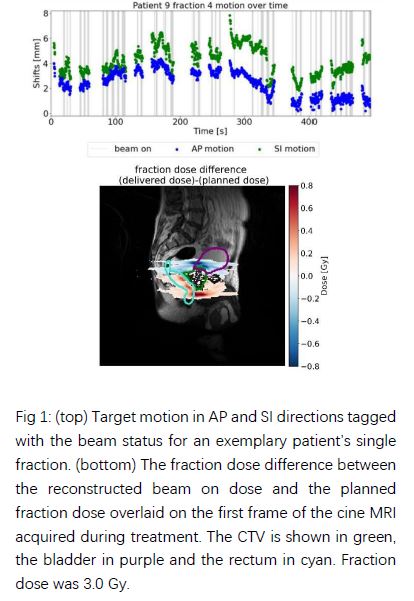
In few single fractions, with pronounced
intra-fractional motion, substantial deviations of reconstructed and planned fraction
dose were observed (Fig. 1). CTV D98% decreased by up to 7%, rectum
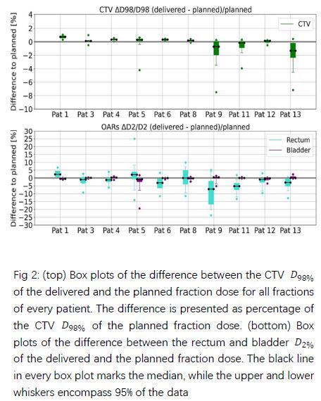
and bladder D2% increased by up to 25% and 3%. However, averaged
over all treatment fractions, CTV D98% showed a decrease within 2% in all patients.
The rectum and the bladder
increase was on average below 3% and 0.5%,
respectively (Fig. 2).
Conclusion
A workflow for extraction of the prostate motion
during MR-guided radiotherapy based on 2D cine-MRI has been implemented. The
obtained motion data can support the estimation of treatment efficiency in
different margin/gating window scenarios and enable reconstruction of the
delivered dose using a dose cloud approximation in future studies. On average,
only minor deviations in target and organs-at-risk dose parameters were
observed, indicating safety of the currently adopted MR-guided treatment
workflow.