Volumetric change in gray matter after radiotherapy in glioma patients detected with MRI
Hye In Lee,
Korea Republic of
MO-0726
Abstract
Volumetric change in gray matter after radiotherapy in glioma patients detected with MRI
Authors: Hyein Lee1, Min Kyoung Kang2, Kihwan Hwang3, Chae-Yong Kim3, Yu Jung Kim4, Koung Jin Suh4, Byung Se Choi5, In Ah Kim6, Bum-Sup Jang6
1Seoul National University Hospital, Department of Radiation Oncology, Seoul, Korea Republic of; 2Nowon Eulji Medical Center, Eulji University, Department of Neurology, Seoul, Korea Republic of; 3Seoul National University Bundang Hospital, Department of Neurosurgery, Seongnam, Korea Republic of; 4Seoul National University Bundang Hospital, Department of Internal Medicine, Seongnam, Korea Republic of; 5Seoul National University Bundang Hospital, Department of Radiology, Seongnam, Korea Republic of; 6Seoul National University Bundang Hospital, Department of Radiation Oncology, Seongnam, Korea Republic of
Show Affiliations
Hide Affiliations
Purpose or Objective
Radiotherapy (RT)-induced volume reduction in gray matter is associated with deterioration of brain function. We performed a large-scale quantitative measurement of volumetric change in gray matter and identified potential predictors of severe volume reduction after RT.
Material and Methods
A single
institution cohort of 461 magnetic resonance imaging (MRI) in 105 patients with
glioma treated with postoperative RT was retrospectively
analyzed. Study patients had MRIs at five timepoints (before and 1 month, 6
months, 1 year and 2 years after RT) and some were missing due to follow-up
loss or death. Using FastSurfer – a deep learning based neuroimaging pipeline,
a total of 81 subsites of brain were automatically segmented from longitudinal
MRIs and their volumetric changes were calculated. Each subsite was classified
into 10 functional fields. A linear mixed effects model was performed to identify
potential predictors of volumetric changes, including gender, tumor grade,
tumor location, RT dose, IDH-mutation, MGMT promoter methylation, 1p/19q
co-deletion, progressive disease after RT, adjuvant chemotherapy, and interaction
between patients’ age at RT and time interval after RT. Statistical
significance was evaluated at a p-value <0.05.
Results
The median volumetric
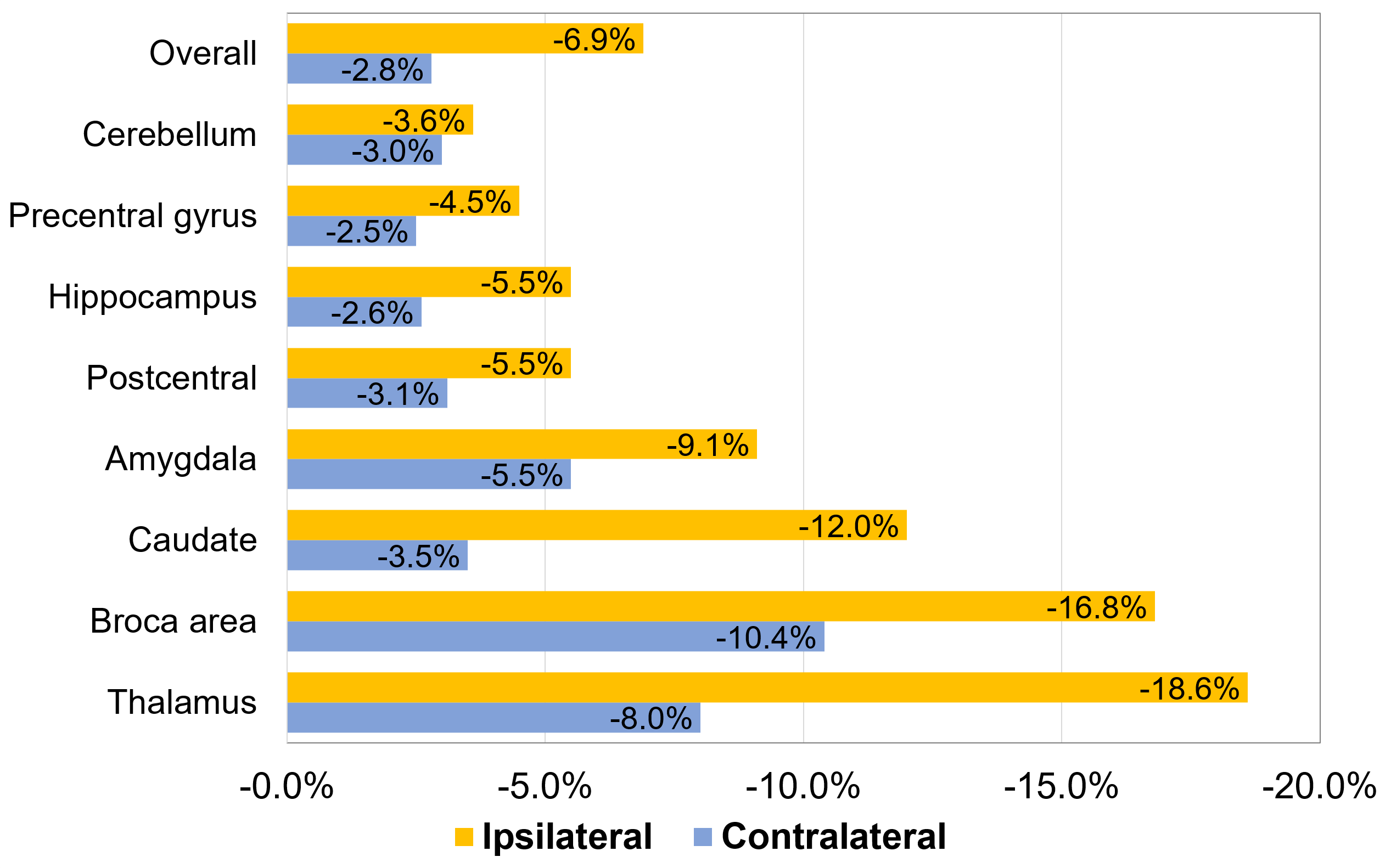
change after RT was -5.1% (range, -37.9% to +4.1%) in overall subsites and the
ipsilateral side with irradiated field showed greater volumetric change than
contralateral side (ipsilateral side: -6.9% [range, -37.8% to +5.3%];
contralateral side: -2.8% [range, -33.3% to +2.6%], p<0.001). The median
volume of hippocampus, caudate, and precentral gyrus were reduced by
5.5%, 12.0% and 4.5%, respectively. In the multivariate analysis, female (vs.
male), longer time interval after RT, older age at RT, and progressive disease
after RT were the best predictors for volume reduction. The interaction between
age at RT and time interval after RT showed significant correlation with
volumetric change, suggesting that the volumetric change over the time after RT
was significant only if the age at RT was older than a specific subsite-dependent
cutoff age (e.g., 45 years for ipsilateral hippocampus and 55 years for ipsilateral
precentral area). Adjuvant chemotherapy and RT dose had little effect on
volumetric change, affecting 4% of subsites. Regarding the functional fields, the areas of
cognitive/emotional, language, and execution of movement showed the largest
volume reduction and the coordinating area (cerebellum) showed the smallest volume
reduction after RT.


Conclusion
Volume reduction in gray matter after RT was observed in most subsites and all functional groups. For the feasible and safe radiotherapy
in brain, the powerful predictors presented in the study should be considered.