Influence of curing time and composition of 3D radiochromic dosimeters on proton LET dependency
Morten Bjørn Jensen,
Denmark
MO-0049
Abstract
Influence of curing time and composition of 3D radiochromic dosimeters on proton LET dependency
Authors: Lia Valdetaro1, Peter Balling2, Peter Sandegaard Skyt3, Mateusz Krzysztof Sitarz3, Jørgen Breede Baltzer Petersen4, Ludvig Paul Muren1
1Aarhus University, Department of Clinical Medicine, Danish Center for Particle Therapy, Aarhus, Denmark; 2Aarhus University, Department of Physics and Astronomy, Interdisciplinary Nanoscience Center, Aarhus, Denmark; 3Aarhus University Hospital, Danish Center for Particle Therapy, Aarhus, Denmark; 4Aarhus University Hospital, Department of Medical Physics, Aarhus, Denmark
Show Affiliations
Hide Affiliations
Purpose or Objective
Silicone-based
radiochromic dosimeters allow for dose read-out in 3D with high spatial
resolution. Additionally, they can be deformed, thus motivating our development
of this dosimeter into a protocol-specific verification tool for several tumour
sites influenced by motion and deformation that will be treated with protons
within controlled clinical trials, including breast and liver cancer. However,
the increase in linear-energy-transfer (LET) with depth can lead to under
response near the Bragg peak. Since the dosimetric properties of this system are
highly dependent on material composition and curing conditions, we investigated
the impact of these factors on the LET dependency.
Material and Methods
Cuvette-sized dosimeters were fabricated from silicone elastomer, curing
agent (CA), chloroform and leucomalachite green for two compositions, containing 5% or 9% CA. The dosimeters were cured in a light-sealed box at
20oC for 1, 3 or 5 days. Proton
irradiation was conducted with monoenergetic beamlets (80 MeV), and an entrance
dose of 5, 10 and 15 Gy. Solid water (SW) slabs were placed around the
dosimeters, including a 2 cm SW slab as a build-up. Dosimeters
were scanned pre- and post-irradiation with a 1D optical scanner (wavelength:
635nm, 0.2 mm steps), with the change in optical density defined as OD=log10(Ipre/Ipost), where Ipre/post is the laser intensity measured after the cuvette. Monte Carlo (MC)
simulations in TOPAS were used to estimate dose (DMC) and dose averaged LET (LETd) distributions, using an experimentally validated beam model. Finally, dose response (OD/DMC) was estimated with linear regression for the three dose
levels measured at 2 cm depth in the dosimeter and used to find the measured dose (Dmeas). The quantity (1 - Dmeas/ DMC)*100
was chosen to quantify the LET dependency.
Results
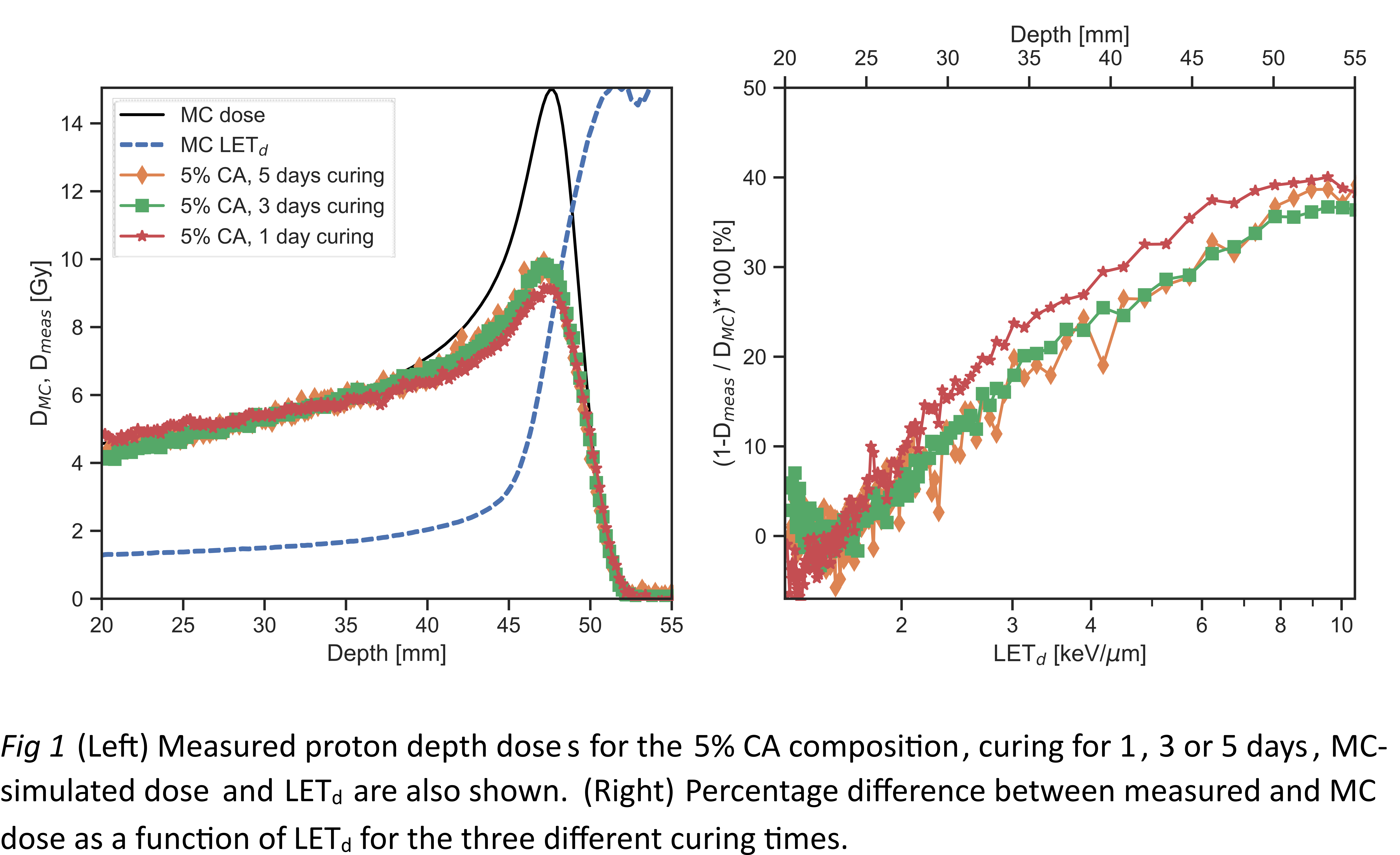
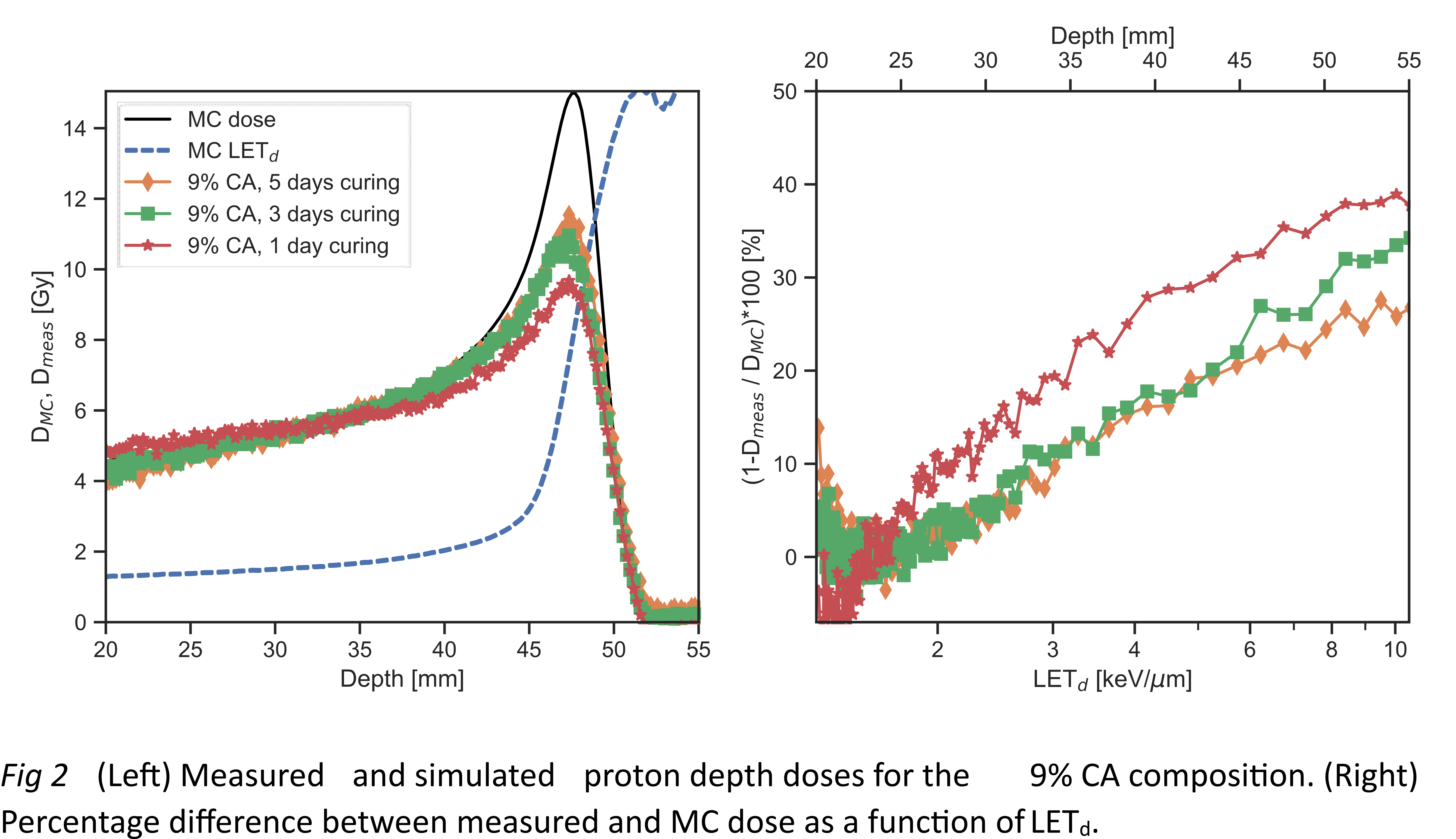
Most of the change happened already after 3 days of curing. For 5% CA dosimeters, LET
dependency decreased from 42% for 1 day of curing (averaged in the LET interval
8.5-10 keV/μm) to 39% for 5
days, whereas for 9% CA it decreased from 41% for 1 day of curing to 26% for 5 days.
The dose response (OD/DMC)
decreased by 13% from 1
to 5 days for 5% CA, and by 34% for 9% CA dosimeters.


Conclusion
Longer curing led to a reduction in LET dependency for both compositions.
For the 5% CA, the reduction was minimal, and coupled with the decrease in dose
response, there was no advantage in extending the curing process. For the 9% CA
we also observed a decrease in dose response, but the significant reduction in
LET dependency (more than 20%) could still be an advantage for some applications.
Regardless, it will always be necessary to correct for the LET dependency of
both dosimeter compositions.