An artificial intelligence approach to tumor volume delineation in glioblastoma
Marianne Hannisdal,
Norway
OC-0462
Abstract
An artificial intelligence approach to tumor volume delineation in glioblastoma
Authors: Marianne Hannisdal1
1Haukeland University Hospital, Department of Oncology and Medical Physics, Bergen, Norway
Show Affiliations
Hide Affiliations
Purpose or Objective
For glioblastoma (GBM), an isotropic margin of
20 mm from the contrast enhancing tumor is used because (i) conventional MRI
and interpretation methods have limitations as to specify the location of
non-enhancing infiltrative GBM, and (ii) recurrence commonly occur within 20 mm
from the primary tumor. However, inter- and intra-observer variability and the
diffuse radiological signature of GBM adds to the challenge of optimal visual
interpretation and accurate delineation of GBM. A precise method is needed to
specify the radiotherapy target and avoid unnecessary patient neurocognitive
side effects.
The objective of this study was to investigate if a deep learning
model for segmenting GBM on multispectral MRI correlated to manual
delineations, and thereby potentially be feasible as an oncologist support
tool. Secondly, we aim to investigate if the 20mm margin is justifiable in
respect of geometrical appearance of recurrent GBM harboring unmethylated O6-methylguanine-DNA
methyltransferase (MGMT) promotor.
Material and Methods
Longitudinal image sets of multispectral
post-operative MRI from six patients with (i) primary- and (ii) recurrent GBM
were analyzed. All patients harbored
unmethylated MGMT promoter, indicating high radioresistance and poor
prognosis. We used a deep learning pre-trained U-Net based algorithm called
HD-GLIO, trained on 3220 GBM image sets labeled by neuro-radiologists. The
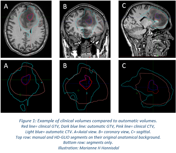
enhancing core (automatic GTV) and non-enhancing GBM compartments (automatic CTV) were derived and compared to the clinical GTVs and CTVs, respectively. The
image series of the primary tumor was rigidly co-registered with the image
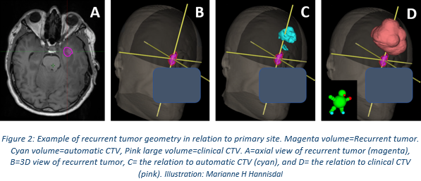
series of the recurrent GBM for volumetric comparison between the clinical CTV and
the recurrent tumor.
Results
The
automatic GTV overlapped with the clinical (true positive) by overall median 90%
(range=33-100%) (p < 0.005), with a mean size=43% compared to the clinical
GTVs. Median Dice coefficient=65% (range=15-77%) (p < 0.005).
The automatic CTV-segments
overlapped with the clinical (true positive result) by median=98% (range=93-100%),
mean size=56% compared to the clinical CTV. Median Dice coefficient=59% (range
15-77%) (p < 0.05).

The recurrent GBM overlapped with the primary clinical
CTV by median 0% true positive (range=0-79%), and median 0% Dice coefficient (range
0-13%) (p < 0, 5).

Conclusion
We found a high degree of overlap in detecting
the spatial area of malignant tissue using HD-GLIO, demonstrating the method feasible
to employ as a support tool for oncologists.
Based on these findings, we have initialized a more extensive study.
We
found a low degree of overlap between CTV-margin and recurrent tumor. This
could suggest radiation treatment response, but considering the known tumor radioresistance it could also mean that irradiating this large margin of potentially normal
tissue cells was only contributing to side effects. We suggest more research is
performed to investigate recurrence patterns in relation to irradiated tissue
volumes on this patient group.