Deep learning and atlas-based approaches for Total Marrow and Lymphoid Irradiation segmentation
PO-1645
Abstract
Deep learning and atlas-based approaches for Total Marrow and Lymphoid Irradiation segmentation
Authors: Damiano Dei1, Nicola Lambri1, Leonardo Crespi2, Ricardo Coimbra Brosio2, Daniele Loiacono2, Marta Scorsetti3, Pietro Mancosu4
1IRCCS Humanitas Research Hospital, Humanitas University, Radiotherapy, Rozzano, Milano, Italy; 2Politecnico di Milano, Dipartimento di Elettronica, Informazione e Bioingegneria, Milano, Italy; 3IRCCS Humanitas Research Hospital, Humanitas University, Radiotherapy, Milano, Italy; 4IRCCS Humanitas Research Hospital, Medical Physics Unit, Rozzano, Milano, Italy
Show Affiliations
Hide Affiliations
Purpose or Objective
Lymphoid clinical target volume (CTV_LN) and organs-at-risk (OARs) delineation in Total Marrow and Lymphoid Irradiation (TMLI) is time-consuming and susceptible to high inter-observer variability. Deep learning (DL) and atlas based (AB) auto-contouring methods have been demonstrated to streamline the RT contouring process for localized treatment sites. However, the use of such algorithms for whole body treatments such as TMLI is still limited. In this study, we investigated DL and AB image segmentation models recently introduced in our clinic to improve the TMLI workflow.
Material and Methods
Ten TMLI patients treated at out Institute were retrospectively selected. A commercial software for DL and AB image segmentation was used (RaySearch Laboratories AB, Stockholm, Sweden). The CTV_LN was segmented using an AB model trained on 20 TMLI patients specifically contoured for this study by a senior radiation oncologist (RO). Twenty-five OARs were segmented using 4 site-specific DL models. TMLI plans were optimized with VMAT and the dose computed for all structures to assess target coverage and OARs sparing. Manual and automatic contours were compared in terms of geometrical agreement, i.e., Dice Similarity Coefficient (DSC) and Hausdorff distance (HD), dose statistics (D98%, Dmean), and time required for segmentation.
Results
Dose statistics revealed clinical differences between manual and AB contours of CTV_LN for D98% (101±2% vs 93±12%), but not for Dmean (110±1% vs 109±2%) (see table 1)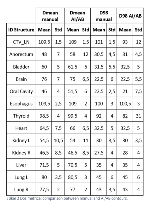 . Good geometrical agreement was found for the majority of OARs, with DSC>=0.93 and HD range (1.17-3.26mm) (see table 2)
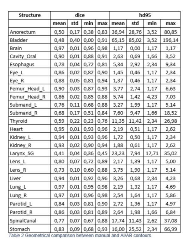 . The most complicated anatomical regions to segment were the larynx, bladder, and rectum, with a DSC of 0.41±0.04, 0.48±0.40, and 0.50±0.20, respectively, and HD of 23±8 mm, 65±85 mm, and 37±29 mm. The AB and DL models generated CTV_LN contours in 8.4±0.6 min, and OARs in 4.3±0.2 min. Since Sept. 2022, all TMLI patients at our Institute are pre-segmented using DL and AB models; the RO reviews and manually adjust the CTV_LN and OARs, with a time-saving >50% with respect to previous manual contouring procedure.
Conclusion
DL and AB models can streamline the RT process for TMLI treatments. A hybrid approach, where the human operator reviews and modifies structures after automatic generation remains fundamental, in particular for CTV_LN delineation.
This work was supported by grant GR-2019-12370739 .