Vienna, Austria

ESTRO 2023

Session Item

Automation
6028
Poster (Digital)
Physics
Autosegmentation of structures for Cranial Spinal Irradiation patients using Deep Learning
Jesper Kallehauge, Denmark
PO-1639

Abstract

Autosegmentation of structures for Cranial Spinal Irradiation patients using Deep Learning
Authors:

Jesper Kallehauge1, Rasin Worawongsakul2, Ole Nørrevang1, Klaus Seiersen1, Yasmin Lassen-Ramshad1

1Danish Centre for Particle Therapy, Aarhus University Hospital, Aarhus, Denmark; 2Ramathibodi Hospital, Mahidol University, Ratchathewi, Department of Diagnostic and Therapeutic Radiology, Bangkok, Thailand

Show Affiliations
Purpose or Objective

Craniospinal irradiation (CSI) is indicated for embryonal brain tumours and leptomeningeal metastasis.  Contouring of target and normal tissue structures for CSI is cumbersome and requires high level expertise to ensure favorable outcomes. The delineation process may easily take more than a day's work even for an experienced physician, however Deep Learning has the potential to significantly reduce the time spent on this process and at the same time improve consistency. The aim of this study was to evaluate the performance of a convolutional neural network (CNN) for segmentation of target and normal tissue organs for CSI.

Material and Methods

Twenty-two individual patient scans were delineated according to the International Pediatric Oncology Society (SIOP) guidelines and used to train and validated four consecutive CNNs while twelve individual patient scans were used for independent testing. Thirty-three individual structures were used for training of which thirteen were related to target and the remaining to normal tissue structures. The performance was evaluated using Dice similarity score (DSC) and the 95th percentile Haussdorff distance (HD95). (Figure 1)  

Results

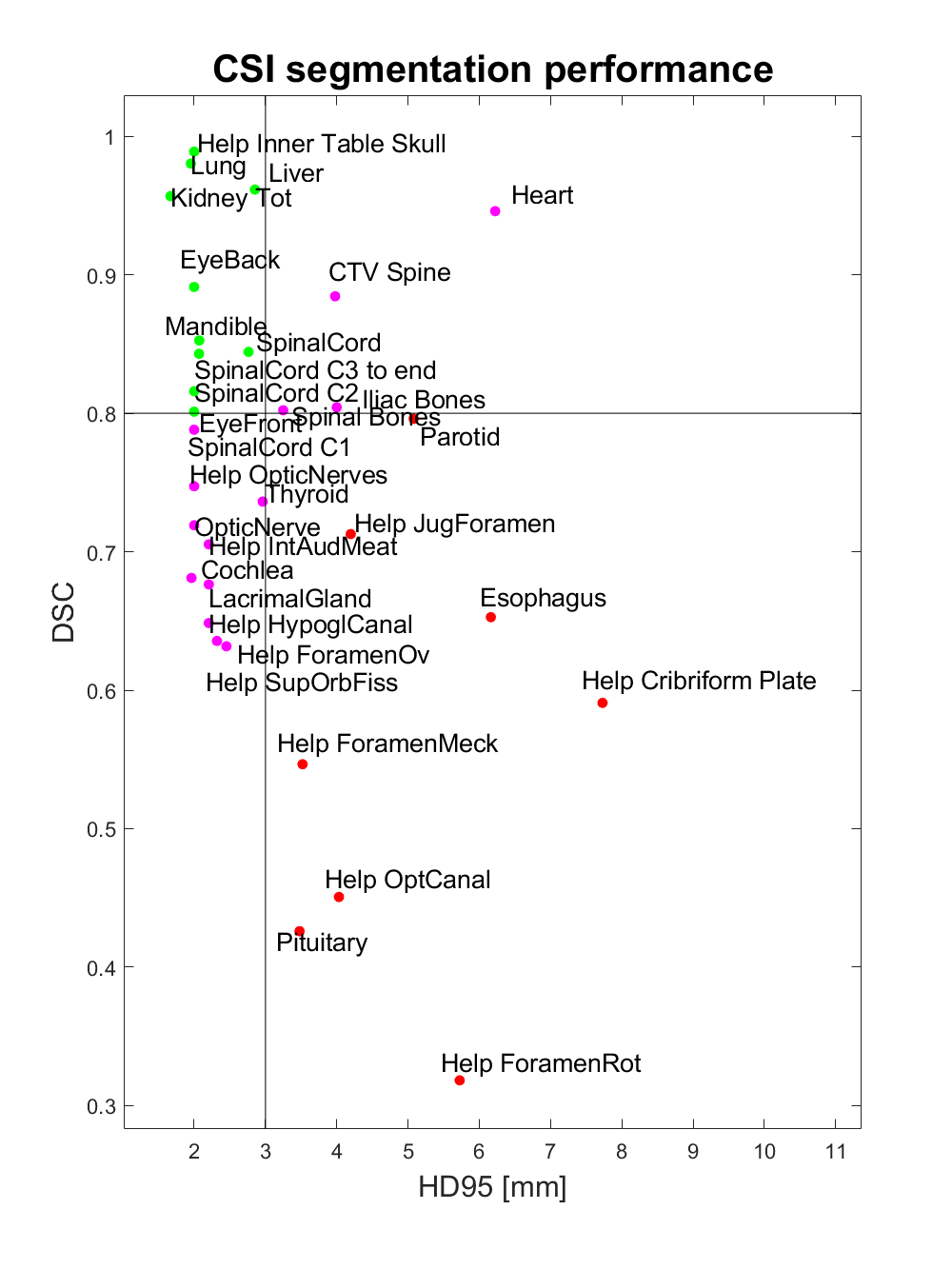
Of the thirty-three structures evaluated, ten structures had a DSC above 0.8 and at the same time a HD95 below 3mm (EyeBack_LR, EyeFront_LR, Kidney_LR, Liver, Mandible, SpinalCord, Lung_LR, SpinalCord_C2, SpinalCord_C3 to end, Help_Inner Table Skull). Eight structures had DSC below 0.8 and HD95 above 3 mm (Parotid_LR, Esophagus, Pituitary, Help_Cribriform Plate, Help_ForamenMeck, Help_ForamenRot, Help_JugForamen, Help_OptCanal). The remaining fifteen structures had either a DSC below 0.8 or a HD95 above 3 mm (Cochlea_LR, LacrimalGland_LR, OpticNerve_LR, Heart, Iliac Bones, Thyroid, Lens_LR, SpinalCord_C1, CTV_Spine, Help_ForamenOv, Help_HypoglCanal, Help_IntAudMeat, Help_OpticNerves, Help_SupOrbFiss, Spinal Boes). (Figure 2)

Conclusion

With the relative limited training set, of twenty-two patient scans available, we saw a good performance in ten annotations related to brain, eyes, mandible, spinal cord, liver and kidneys while we saw poor prediction quality mainly related to target help structures in the brain. The remaining structure had mediocre prediction quality. Including more patient scans into the training set would very likely improve the inductive bias of the model, but already with the current presented results we can identify relevant structures that are of high, mediocre and poor quality. This can guide us towards where the model requires improvement and in a clinical implementation what segmentations to be more or less trustworthy of.