Reproducibility of AI-based contour generation on synthetic CT
PO-1625
Abstract
Reproducibility of AI-based contour generation on synthetic CT
Authors: Ivan Coric1, Nicole Wernlein1, Marcel Nachbar1, Daniel Wegener2, Simon Boeke2, Daniel Zips2, Cihan Gani2, Daniela Thorwarth1
1Section for Biomedical Physics. Department of Radiation Oncology, University Hospital and Medical Faculty. Eberhard Karls University Tuebingen, Tuebingen, Germany; 2Department of Radiation Oncology, University Hospital and Medical Faculty. Eberhard Karls University Tuebingen, Tuebingen, Germany
Show Affiliations
Hide Affiliations
Purpose or Objective
Automatic segmentation based on artificial intelligence (AI) offers a time-saving solution for delineation of organs at risk (OARs), which is essential for an online adaptive workflow at the MR-Linac. With the introduction of synthetic CTs (sCT) in radiotherapy, the need for registration of a planning CT to the daily MRI can be bypassed. However, the use of sCTs can be extended for delineation of OARs in the daily adaptation routine. While the segmentation of bony structures is dosimetrically relevant but proves to be difficult on MRI, the sCT provides remediation. In this work, the reproducibility of OAR segmentations generated by AI-based MRI and CT delineation algorithms on MRI and sCT was compared.
Material and Methods
A total of 30 primary prostate cancer patients treated on the MR-Linac were included. For each patient, sCTs were generated from T2w-MRIs using a generative adversarial neural network (GAN). OARs were automatically contoured on the T2w-MRI and sCT using ART-Plan™ (TheraPanacea, Paris, France), respectively. MRIs were delineated by an AI-based MR segmentation method, which was trained on 235 T2w-MRIs. For the sCTs, a commercial, pretrained AI-based CT segmentation algorithm was used. The structures anal canal, bladder, femur left/right and rectum were considered for evaluation. Comparison was performed by calculating Hausdorff Distance (HD), 95% percentile HD (HD95), volumetric Dice similarity coefficient (vDSC), surface DSC (sDSC), path length (PL) and added path length (APL) using MATLAB (R2020a).
Results
Results were successfully generated for 28/30 data sets. Lowest median HD and HD95 were observed for anal canal (HD=7.2 mm) and bladder (HD95=2.0 mm). In terms of median vDSC, bladder showed the highest agreement with 0.97, while lowest agreement was observed for anal canal with 0.71. Median sDSC ranged from 0.48 for anal canal to 0.94 for rectum. The lowest median APL was determined for anal canal with 57.4 cm.
Table 1 Comparison of the investigated structures for each calculated metric. Shown is the median [range] value of the respective metric.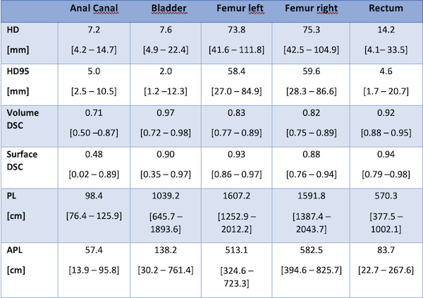
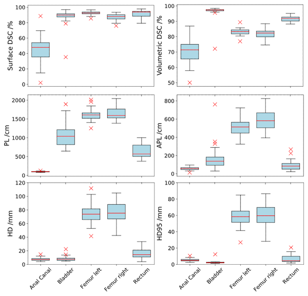
Figure 1 Visualization of the calculated metrics for each evaluated structure.
Conclusion
Detailed analysis of AI-based segmentation for two different AI-algorithms on different image modalities demonstrated in comparison acceptable deviations. While the auto-segmentation of bladder and rectum was reproduced correctly for most of the patients, anal canal showed lower agreement regarding vDSC and sDSC. Details may also be due to differences in the training contours of the used MRI and CT delineation algorithms. As the MRI provides superior soft tissue contrast for delineation of target volumes and OARs, the use of sCT for automatic segmentation of bony structures appears reasonable for an online MRgRT workflow.