Vienna, Austria

ESTRO 2023

Session Item

Automation
6028
Poster (Digital)
Physics
Evaluation of a CT scanner based deep learning auto-contouring solution for lung radiotherapy
Matthew Williams, United Kingdom
PO-1622

Abstract

Evaluation of a CT scanner based deep learning auto-contouring solution for lung radiotherapy
Authors:

Matthew Williams1, Salvatore Berenato1, Christian Möhler2, Anthony Millin1, Philip Wheeler1

1Velindre Cancer Centre, Radiotherapy Physics, Cardiff, United Kingdom; 2Siemens Healthineers, ,, Forchheim, Germany

Show Affiliations
Purpose or Objective

For non-small cell lung cancer radiotherapy (NSCLC), geometrically and dosimetrically evaluate DirectORGANS (Version VA30): a commercial AI solution that is natively integrated into a CT scanner and utilises dedicated reconstructions optimised for auto-contouring.

Material and Methods

CT scans of 20 NSCLC patients were sequentially selected to evaluate AI contours for lungs (lt & rt), heart, oesophagus (oes) and cord. 3 plan generation ‘pipelines’ were considered; the use of AI generated contours (AI-Std), full amendment of the AI generated contours (AI-FullEd), and manual delineation MD-Ob1. Contouring time was recorded and plans generated for each pipeline using a validated 55Gy in 20# automated planning solution.

Contour sets AI-Std and AI-FullEd were geometrically and dosimetrically compared to MD-Ob1. For dosimetric comparison the error in both Reported Dose (RD) and Patient Dose (PD) was evaluated. RD was defined as DVH parameters that would be reported in patient records for a given pipeline. The dose distribution of each pipeline plan was evaluated on both the reference (RD-Ref) and pipeline (RD-Pipe) contour sets, with the difference calculated to assess the impact of contour discrepancies on RD. PD was defined as the best estimate of the actual dose the patient would receive and was extracted from the pipeline plan’s DVH using the MD-Ob1 (PD-Pipe). By comparing PD-Pipe with plans generated by and evaluated using MD-Ob1 (PD-Ref), a contour set’s influence on the optimisation process and hence final dose distribution, was assessed.

Results

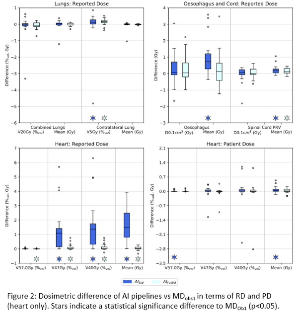
Compared to MD-Ob1, AI-FullEd reduced median delineation time by 50%, 61%, 37%, 28% and 14% for lung_lt, lung_rt, oes, cord and heart respectively. For heart, AI-FullEd increased delineation time in 8 of 20 cases. For lung_lt, lung_rt, oes and cord, AI-Std contours exhibited good geometric alignment to MD-Ob1 with median mean surface distances (MSD) <1.1mm and median DSC results of 0.97, 0.98, 0.80 and 0.88 respectively. For heart, agreement was poorer (MSD=3.4mm, DSC=0.90).  AI-FullEd led to small improvements in overall agreement for lung, cord and oes, with moderate improvements for heart (Fig1).


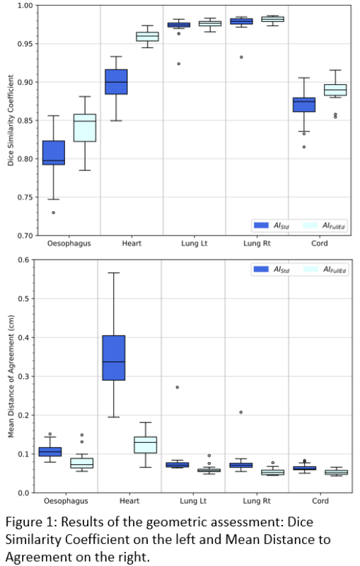
Except for heart RD, all pipelines exhibited excellent dosimetric agreement with MD-Ob1 (Fig2). For RD, median deviations were within ±0.1% (c.f. ±1.4% for heart) and ±0.7Gy (c.f. ±1.5Gy) for relative volume and dose metrics respectively. For PD, agreement was improved with respective values (inc. heart) within ±0.2% and 0.1Gy. Except for a few outliers and heart RD, the distribution of PD/RD deviations for AI-Std and AI-FullEd was considered nominally equivalent.

Conclusion

Whilst the heart model could be improved, AI contours demonstrated good geometric agreement with MD-Ob1 and reduced delineation time. Dosimetric analysis showed that except for gross outliers and heart RD, the effect of AI-Std contour variation on RD and PD was nominally equivalent to AI-FullEd. AI-Std could therefore be clinically implemented with no degradation in plan dosimetry.