Evaluation of commercial deep learning MRI reconstruction for synthetic CT generation in prostate RT
Christian Jamtheim Gustafsson,
Sweden
PD-0979
Abstract
Evaluation of commercial deep learning MRI reconstruction for synthetic CT generation in prostate RT
Authors: Christian Jamtheim Gustafsson1,2, Jonas Scherman1, Adalsteinn Gunnlaugsson1, Lars E Olsson3
1Skåne University Hospital, Dept Haematology, Oncology and Radiation Physics, Lund, Sweden; 2Medical Radiation Physics, Lund University, Department of Translational Sciences, Malmö, Sweden; 3Medical Radiation Physics, Lund University , Department of Translational Sciences Malmö, Malmö, Sweden
Show Affiliations
Hide Affiliations
Purpose or Objective
The use of deep learning (DL) for magnetic resonance image (MRI) reconstruction has been implemented by several commercial vendors and allows for both image noise- and scan time reduction. This can provide several benefits when using MRI for radiotherapy planning purposes. The aim of this work was 1) to evaluate the compatibility between a commercial DL MRI reconstruction product and a commercial synthetic CT (sCT) generation software and 2) to quantitatively assess the Hounsfield (HU) and dosimetric integrity of sCT created from such MR images.
Material and Methods
Twenty-four prostate cancer patients were prescribed ultra hypofractionated radiation therapy (RT) with 42.7 Gy, 7 fractions, in a clinical MRI-only treatment workflow using a General Electric (GE, Chicago, USA) 3T Architect MRI system together with a lightweight AIR receiver coil. Within the clinical MRI acquisition protocol, a large field of view (LFOV) T2 weighted MRI image volume was acquired for sCT conversion (sCT_orig) using Spectronic MRI planner v.2.4.14 (Spectronic Medical AB, Helsingborg, Sweden). This sCT generation has previously been validated against CT. In parallel, the MRI raw data from the LFOV acquisition was used in DL MRI reconstruction using the GE Air Recon DL product for host version MR29.1 and a new MRI image volume was created together with a corresponding sCT (sCT_DL).
To assess differences in HU values between sCT_orig and sCT_DL, HU differences outside the patient, in fat, muscle, spongy bone and compact bone was analyzed. To assess RT dose differences in target and organs at risk, clinical RT structures except the body structure were copied from the sCT_orig to the sCT_DL, clinical treatment plan was transferred with the same number of monitor units and dose was recalculated on sCT_DL. The created sCT_DL was visually inspected and the body volume for all sCT were calculated.
Results
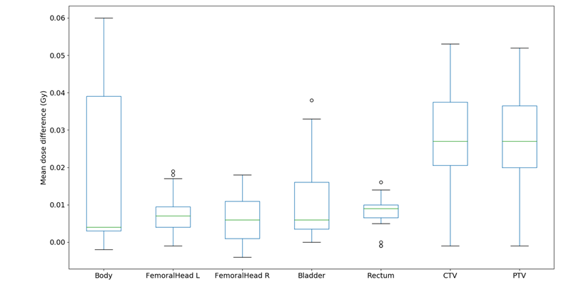
HU differences outside the patient, in fat, muscle, spongy bone and compact bone had a mean absolute error (± 1 STD) in the cohort of 1.5±0.4, 1.5±0.3, 1.2±0.4, 4.9±1.1 and 7.4±2.2 HU (n=24), respectively. The mean dose differences for body, femoral heads, bladder, rectum, CTV and PTV were all positive and below 0.06 Gy (Fig.1, 0.1% difference, n=23). The sCT_DLs had a visually excepted appearance and the sCT body volume were larger for all patients but three compared to sCT_orig, with a median cohort difference of 15 cm3 (spread in cohort body volume was 11603-21542 cm3).

Fig.1. Mean dose differences for OAR and target (sCT_orig-sCT_DL). A small systematic shift towards a positive dose difference was observed. This was probably due to the larger sCT_DL volume, providing more radiation attenuation.
Conclusion
DL MRI reconstruction was suitable for sCT generation with only minor, clinically negligible, differences in HU and calculated dose. The differences were systematic and probably due to the larger size of the sCT_DL, and believed to be the effect of improved MRI image quality, thereby affecting the sCT generation.