Cardiac SABR: image matching techniques for accurate treatment delivery
Rachel Brooks-Pearson,
United Kingdom
OC-0939
Abstract
Cardiac SABR: image matching techniques for accurate treatment delivery
Authors: Rachel Brooks-Pearson1, Karen Pilling1, Bethany Ormston1, Laura MacKenzie1, Claire Huntley2, Alison Kerr2, Rosaleen Crouch3, Neil Richmond1, Marieke Van Der Putten1, Philip Atherton1
1Newcastle upon Tyne Hospitals NHS Foundation Trust, Northern Centre for Cancer Care, Newcastle upon Tyne, United Kingdom; 2South Tees Hospitals NHS Foundation Trust, Radiotherapy, James Cook University Hospital, Middlesbrough, United Kingdom; 3Sheffield Teaching Hospitals NHS Foundation Trust, Radiotherapy, Weston Park Hospital, Sheffield, United Kingdom
Show Affiliations
Hide Affiliations
Purpose or Objective
Cardiac SABR is a novel technique to treat non-malignant heart disease. Ventricular tachycardia is an irregular heartbeat conventionally treated using invasive cardiac catheter ablation and medication. However, when these standard treatments have been exhausted, cardiac SABR provides a final treatment option to this high-mortality condition.
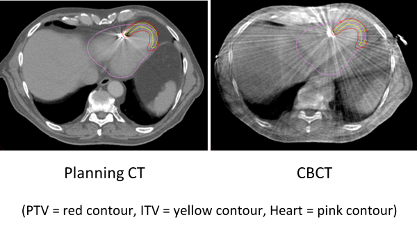
Complex diagnostic mapping and planning scans enable the cardiologist and clinical oncologist to delineate the target for a 25Gy single fraction. However, organs at risk (OAR) near the target make this treatment challenging to plan and deliver. The image below shows planning volumes and image quality of the planning CT compared to the treatment CBCT. Publications from cardiologists report the efficacy of cardiac SABR, however there is limited data on the treatment delivery of this complex procedure. To the best of our knowledge, this is the first study to investigate radiographer CBCT image matching techniques for cardiac SABR.

Material and Methods
Four specialist therapeutic radiographers reviewed 40 CBCTs from the first 10 patients treated in the UK. The 4 radiographers all had cardiac anatomy training from a radiologist and had experience in delivering cardiac SABR treatment. Anonymised images were imported into a Varian training system from 3 UK centres using CBCTs from both Elekta and Varian linacs. A series of image matches were conducted by each radiographer: a manual match (manual), an automatic match to the heart structure (auto) and the auto match followed by manual adjustment to the PTV (PTV), all using three degrees of freedom (DoF) only. The auto and PTV matches were also repeated using 6 DoF. Inter-observer variability was quantified using 95% limits of agreement from a modified Bland-Altman analysis.
Results
Table 1. Limits of agreement (mm)
| (mm) | Lateral | Vertical | Longitudinal | Pitch | Yaw | Roll |
| 3DoF Manual | 1.69 | 1.87 | 2.03 | - | - | - |
| 3DoF Auto | 0.94 | 1.05 | 1.40 | - | - | - |
| 3DoF PTV | 1.57 | 2.06 | 2.11 | - | - | - |
| 6Dof Auto | 0.85 | 0.99 | 1.06 | 0.58 | 0.38 | 0.52 |
| 6DoF PTV | 1.06 | 1.24 | 1.68 | 0.65 | 0.44 | 0.51 |
The table shows the limits of agreement were smallest in the automatic matches which suggests the algorithm matching to the heart structure is reliable. A manual adjustment from the auto match to the PTV is clinically appropriate to optimise target coverage. The mean difference between the 3DoF PTV match and the 6DoF PTV match is 0.6 mm, which could be clinically significant in a target with small margins and OAR in close proximity to high dose gradients. The limits of agreement were smaller in the 6DoF auto and PTV matches than the 3DoF matches. This demonstrates that when the rotations are corrected for as part of the match, matching to the PTV is more consistent.
Conclusion
This data shows that a 6DoF CBCT image match has less variability and therefore more accurate treatment delivery. Therapeutic radiographers have no clinical experience of matching to a target in the heart, this multi-centre study addresses the gap in the knowledge base.