Scintillation imaging for time-resolved 2D monitoring of ultra-high dose rate proton beam scanning
OC-0929
Abstract
Scintillation imaging for time-resolved 2D monitoring of ultra-high dose rate proton beam scanning
Authors: Eleni Kanouta1, Petr Bruza2, Megan Clark2, Jacob Sunnerberg2, Joseph Harms3, Jacob Johansen1, Per Poulsen1
1Aarhus University Hospital, Danish Centre for Particle Therapy, Aarhus, Denmark; 2Dartmouth College, Thayer School of Engineering, Hanover, NH, USA; 3University of Alabama at Birmingham, Department of Radiation Oncology, Birmingham, AL, USA
Show Affiliations
Hide Affiliations
Purpose or Objective
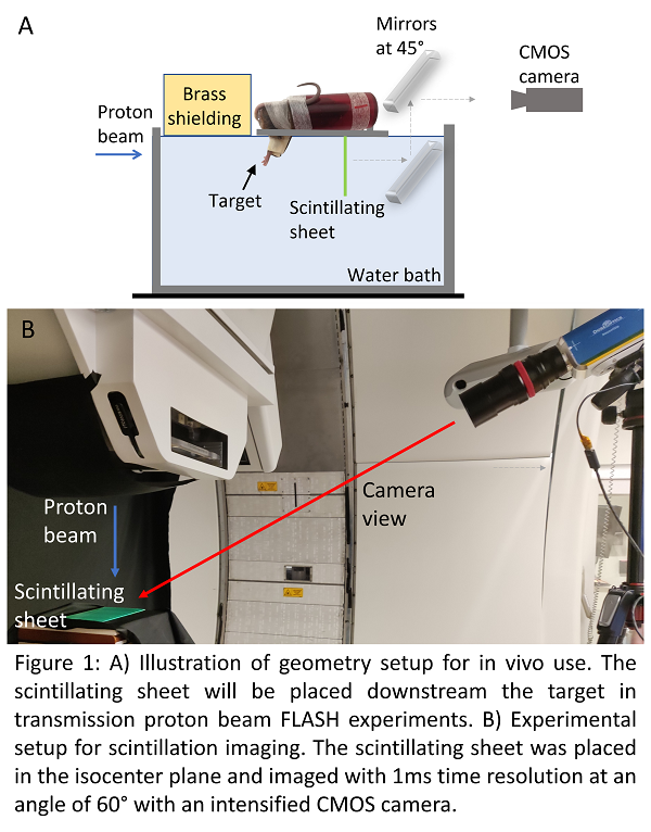
Dosimetry is essential for FLASH radiotherapy studies. It requires dosimeters capable of handling ultra-high dose rates (UHDR). For pencil beam scanning (PBS) proton beams, high spatial and temporal resolution is furthermore required to provide information about each individual spot delivery. Here we propose a novel high-speed camera-based, time-resolved 2D in vivo dosimetry, where a transparent scintillator sheet placed downstream the target in transmission proton FLASH experiments would allow direct monitoring of the dose and dose rate distribution relative to the target anatomy (Fig. 1A). In this first study we characterize the high-speed camera and transparent scintillator sheet with UHDR PBS proton beams.
Material and Methods
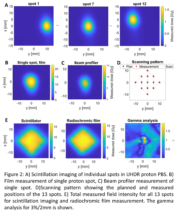
A 1-mm thick transparent scintillator (peak emission wavelength 490nm) sheet in the isocenter plane was imaged using a fast intensified CMOS camera, located 2m from the isocenter at 60° angle from the sheet (Fig. 1B). The sheet was imaged at 1kHz rate with 1µs dead time, which enabled imaging of individual spots. A 2x2 cm2 UHDR PBS field with 13 spots was delivered using a 250MeV proton beam and 99nA beam current (320Gy/s at the spot center). The raw images were background subtracted, flat field corrected, spatially filtered with a Gaussian filter and geometrically transformed from the camera view to a beams’ eye view. The profile of each spot was determined by averaging the intensity across all frames in a spot position. The measured total field intensity was found by summing the intensity of all frames. The summed images for both the field and single spots were calibrated with a PPC05 ionization chamber and compared to radiochromic film measurements as well as a commercial scintillation-based beam profiler.
Results
The system resolved all individual beam spots temporally (Fig. 2A) with 0.1mm pixel and 0.6mm spatial resolution. The measured signal intensity fluctuated 3% between spots. The Gaussian width of individual spots in the scintillation images was 0.6mm smaller than the width measured with a radiochromic film (Fig. 2B), while it matched better with the reference scintillation-based profile (Fig. 2C), confirming the known film UHDR overresponse. For the scanning field (Fig. 2D), the spot center locations matched the plan within 0.3 and 0.1mm in x and y direction, respectively. The field at 50% isodose showed an average difference of 1.5mm between the scintillator and the film (Fig. 2E).
Conclusion
Scintillation imaging using a transparent scintillator sheet offers a unique dosimetry method, allowing UHDR dose rate measurements of individual spots. Further work on characterizing the scintillator sheet response at different geometries will allow direct 2D monitoring of the instantaneous dose rate of UHDR proton PBS with sub-millimeter spatial resolution, all with direct spatial correlation to the target’s external anatomy.