Intrafraction tumor motion monitoring and dose reconstruction for liver spot scanning proton therapy
PD-0250
Abstract
Intrafraction tumor motion monitoring and dose reconstruction for liver spot scanning proton therapy
Authors: Saber Nankali1,3, Esben Worm2, Jakob Thomsen1, Line Stick1, Morten Høyer1, Britta Weber1,2, Hanna Mortensen1, Per Poulsen1,2
1Aarhus University Hospital, Danish Centre for Particle Therapy, Aarhus, Denmark; 2Aarhus University Hospital, Department of Oncology, Aarhus, Denmark; 3Aarhus University, Department of Clinical Medicine, Aarhus, Denmark
Show Affiliations
Hide Affiliations
Purpose or Objective
To develop intra-treatment tumor motion monitoring during pencil beam scanning (PBS) proton therapy and combine it with motion-including tumor dose reconstruction for individual hepatocellular carcinoma (HCC) treatment fractions.
Material and Methods
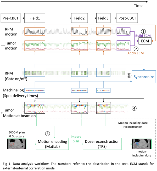
Three patients with HCC were treated with proton PBS using respiratory gating around the exhale phase. A 3-field IMPT plan was made on the exhale phase of a 10-phase 4DCT using an iCTV that encompassed the five 4DCT phases closest to full exhale. The prescribed mean CTV dose was 58 GyRBE (n = 2) or 67.5 GyRBE (n = 1) in 15 fractions. Daily patient setup was based on a CBCT scan in which the exhale positions of three implanted fiducial markers were matched with the planning CT. Throughout the whole treatment session the position of a marker block on the patient’s abdomen was recorded with an optical camera (RPM, Varian). The RPM signal was used during treatment for exhale respiratory gating with a duty cycle of approximately 50%. A post-treatment control CBCT scan was captured at 6, 1 and 7 fractions for patients 1, 2 and 3, respectively. After treatment the fiducial markers were segmented in all raw 2D projections of the post-treatment CBCTs. Then, their 3D motion trajectories during the scan were estimated by a probability-based method and used as surrogates for the tumor position. To estimate the tumor motion during treatment delivery the RPM signal was first synchronized with the post-treatment CBCT projections, and an augmented linear external-internal correlation model (ECM) that estimated the tumor motion from the RPM motion was built (Label 1 in Fig 1). Next, the ECM was used to estimate the tumor motion throughout the treatment delivery (Label 2). The unique pattern of gate-on and gate-off periods during field delivery was then used to synchronize the RPM signal with machine log files that contained the delivery time of each spot (label 3). This synchronization resulted in the tumor position at the time of each spot delivery (label 4). Finally, the motion-including fraction dose to the CTV was estimated with a dose reconstruction method that emulates tumor motion in beam’s eye view as lateral spot shifts and in-depth tumor motion as changes in the proton beam energy (label 5). The motion-encoded plans were imported and calculated in the treatment planning system, and the resulting CTV doses were compared with the planned dose.
Results
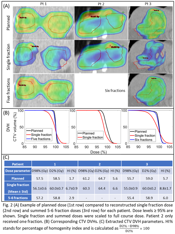
The tumor position during spot delivery had a root-mean-square error of 1.25mm (LR) 2.8mm (CC) 1.7mm (AP) compared to the planned position. Large dose deterioration occurred at single fractions due to interplay effects, but these were to a large degree washed out over 5-6 fractions due to averaging effects (Fig 2).
Conclusion
A method to estimate internal tumor motion and reconstruct the motion-including fraction dose for PBS proton therapy in the liver was developed and implemented successfully in the clinic.

