Can surface imaging predict the impact of anatomical deformations on proton breast treatment?
Vincent Hamming,
The Netherlands
PD-0248
Abstract
Can surface imaging predict the impact of anatomical deformations on proton breast treatment?
Authors: Vincent Hamming1, Daniele Cannavò1, Bojan Strbac1, Gabriel Guterres Marmitt1, John H. Maduro1, Johannes A. Langendijk1, Nanna M. Sijtsema1, Stefan Both1
1University Medical Center Groningen, Radiotherapy, Groningen, The Netherlands
Show Affiliations
Hide Affiliations
Purpose or Objective
Currently proton breast cancer patients receive weekly repeat CTs for treatment robustness evaluation purposes, often showing that the original treatment remains robust. Therefore, the goal of this feasibility study is to assess if breast surface anatomy deformation can predict the need for repeat CT acquisition and plan adaptation in proton breast cancer patients.
Material and Methods
A phantom test and seven proton breast cancer patients with 109 daily Surface Guided RadioTherapy (SGRT) images in treatment position were included in this proof of principle study. The phantom test consisted of imaging (CT and SGRT) a mannequin with known deformations of 5;10;15;20;25 and 35mm. In the Treatment Planning System (TPS), a proton plan was created for this phantom based on a cylindrical target. The plan was subsequently re-calculated on the CT sets with the deformations. Firstly the SGRT images were rigidly registered to the BODY contour from the planning CT by an iterative closest point algorithm employing a novel in-house developed tool. Secondly, a landmark based deformation analysis was performed to determine the local deformation. The magnitude of the local deformation was defined as the ratio of the maximum and the minimum vector length between landmarks, captured in the value T. Hence, T is a unique estimator whose value is close to 1 for similar shapes and increases with deformation present. Within the phantom test the correlation between T and the dose difference with respect to the original CT was determined for the CTV dose-volume parameters D99, D98, D95 and mean dose.
For the clinical patients, deformations of 0 to +20mm were applied to the BODY contour at the location of the target area to determine the sensitivity of the tool and similar analyses were performed as for the phantom.
Results
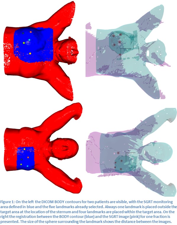
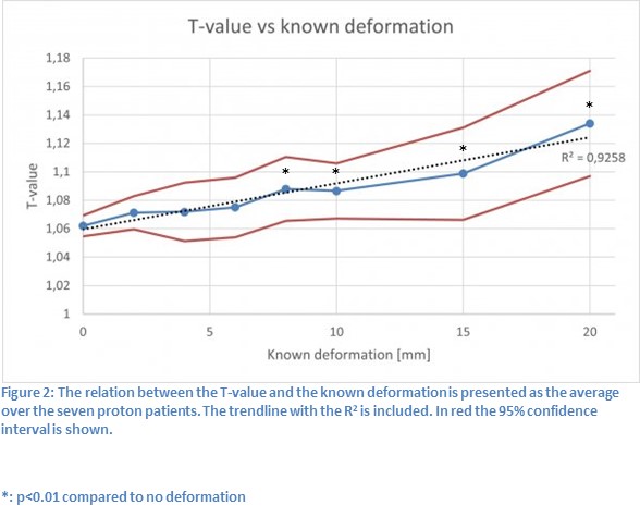
Five landmarks were used, one was placed around the sternum as a reference point, whereas the other four landmarks were placed inside the field of view of the SGRT images and within the target area. Figure 1 shows two examples of the BODY including landmarks, the corresponding registration between the BODY and the SGRT image and the resulting difference in landmark position. For the phantom test, the correlation coefficient of T with the known deformation was 0.91, with the D99, D98 and D95 it was 0.99 and with the mean dose it was 0.82. For the patient group, Figure 2 shows the relation between T and known BODY deformations averaged over the seven proton patients. A known deformation of 8 mm and larger within the patient group showed a significant difference compared to no deformation between the T values.

Conclusion
Breast surface anatomy deformation is highly corelated with target dosimetrical indicators and therefore is potentially a reliable predictor for the need of repeat CT acquisition and plan adaptation in proton breast cancer patients. Future work is warranted to determine accurate thresholds in daily clinical workflow.