Superficial HDR brachytherapy - dosimetric verification of dose distribution with lead shielding
PD-0501
Abstract
Superficial HDR brachytherapy - dosimetric verification of dose distribution with lead shielding
Authors: Grzegorz Bielęda1,2, Grzegorz Zwierzchowski1,2, Agata Szymbor3, Marek Boehlke4
1Greater Poland Cancer Centre, Medical Physics, Poznań, Poland; 2Poznan University of Medical Sciences, Electroradiology, Poznań, Poland; 3MCO Affidea, Medical Physics, Poznań, Poland; 4West Pomeranian Oncology Center, Medical Physics, Szczecin, Poland
Show Affiliations
Hide Affiliations
Purpose or Objective
Surface brachytherapy, usually characterized by a high dose gradient, allows the dose to be precisely deposited in the irradiated area while protecting critical organs. When the lesion is located in the nasal or ocular region, the organ of vision must be protected. The aim of this study was to verify the dose distributions near critical organs in the head and neck region during brachytherapy procedure using lead shielding of the eye using an anthropomorphic phantom and an applicator manufactured from PLA (poli-lactic acid) using FDM (fused deposition modeling) 3D printing technology.
Material and Methods
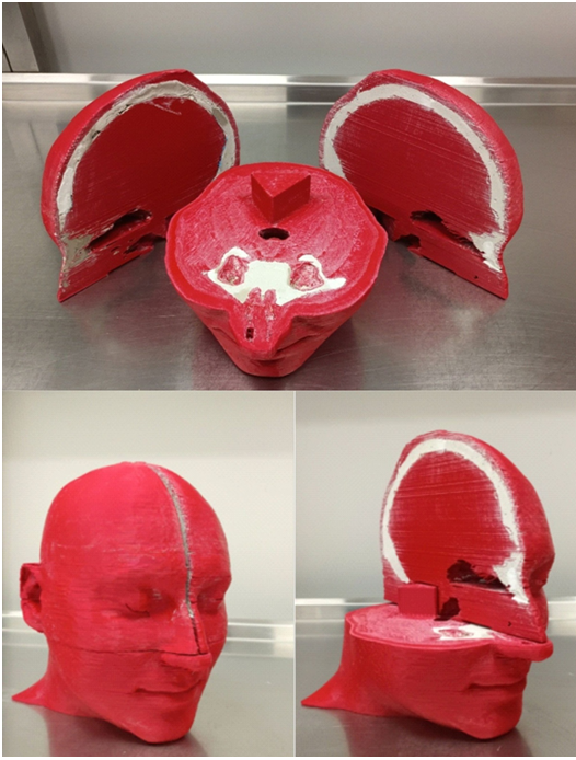
An anthropomorphic head phantom with preserved air spaces and imitation cranial bones filled with plaster of a density comparable to real bones (600-700 HU) was prepared (fig1).

Figure 1. Finished 3D printed anthropomorphic head phantom used for purpose of this study.
Separate treatment plans were prepared for 10 target areas located in different regions of the face. An individual applicator was designed with channels for the source matched to the volume of the PTVs. A space was designed in the applicator for the eye lead shield, and each plan was prepared with a variant with and without the shield. When no shielding was in place a water equivalent dummy shield was inserted. Doses were measured at a point located in the lens of the eye and on the skin surface of the eyelid using EBT3 radiochromic films. Measured doses were compared to doses calculated using TG43 and TG186 formalism algorithms implemented in Oncentra Brachy 4.5.2 TPS.
Results
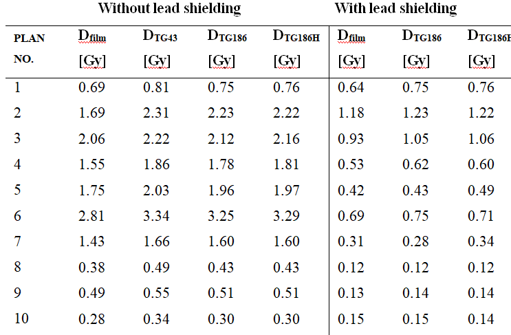
Table 1. Table summarizes doses deposited to lens point measured by radiochromic film and calculated using TPS.

Doses measured by Gafchromic EBT3 films for treatment plans with lead shielding were found to be statistically consistent with the TG186 formalism in 7 out of 10 cases (CTV 2, 5, 6, 7, 8, 9, 10). In the case of CTV 5, however, the TG186H high accuracy mode did not show compliance (p=0.023), for the other plans there were no statistically significant differences observed for the TG186H vs. TG186 mode of the calculation engine. The average readings from the films turned out to be close to the results proposed by TPS and taking into account the presence of lead shielding in 5 out of 10 plans. On the other hand for plans CTV 1, 2, 3, 4, and 6, the dose values measured by the films were lower than those calculated by the treatment planning system.
Conclusion
Doses calculated in the treatment planning system were compared with doses measured using radiochromic films - for the personalized surface brachytherapy applicator. The differences observed are the consequence of the inability of this method to record short-range, low-energy radiation backscattered from the shielding material. It can be assumed that the use of lead shields is a method for protecting the organ of vision from the adverse effects of ionizing radiation.