Technique and oncological outcomes with high dose rate skin surface brachytherapy for skin tumors.
PO-2191
Abstract
Technique and oncological outcomes with high dose rate skin surface brachytherapy for skin tumors.
Authors: Kaoutar SOUSSY1, Mohamed Ait Erraisse1, Mohamed Baraka2, Wissal Hassani1, Fatima Zahraa Farhane1, Zenab Alami1, Touria Bouhafa1
1Hassan II University Hospital, Radiation Oncology Department, Fes, Morocco; 2Hassan II University Hospital, Medical Physics Unit, Fes, Morocco
Show Affiliations
Hide Affiliations
Purpose or Objective
Introduction: High dose rate skin surface brachytherapy (HDR SS-BT) is a useful non-invasive technique for treatment of skin tumours, with good local control rates as well as excellent cosmetic and functional outcomes.
Objective: we report in this study, the experience of the radiation oncology department of the university hospital of Fes Morocco in HDR SS-BT, by describing the technique used and the cases treated.
Material and Methods
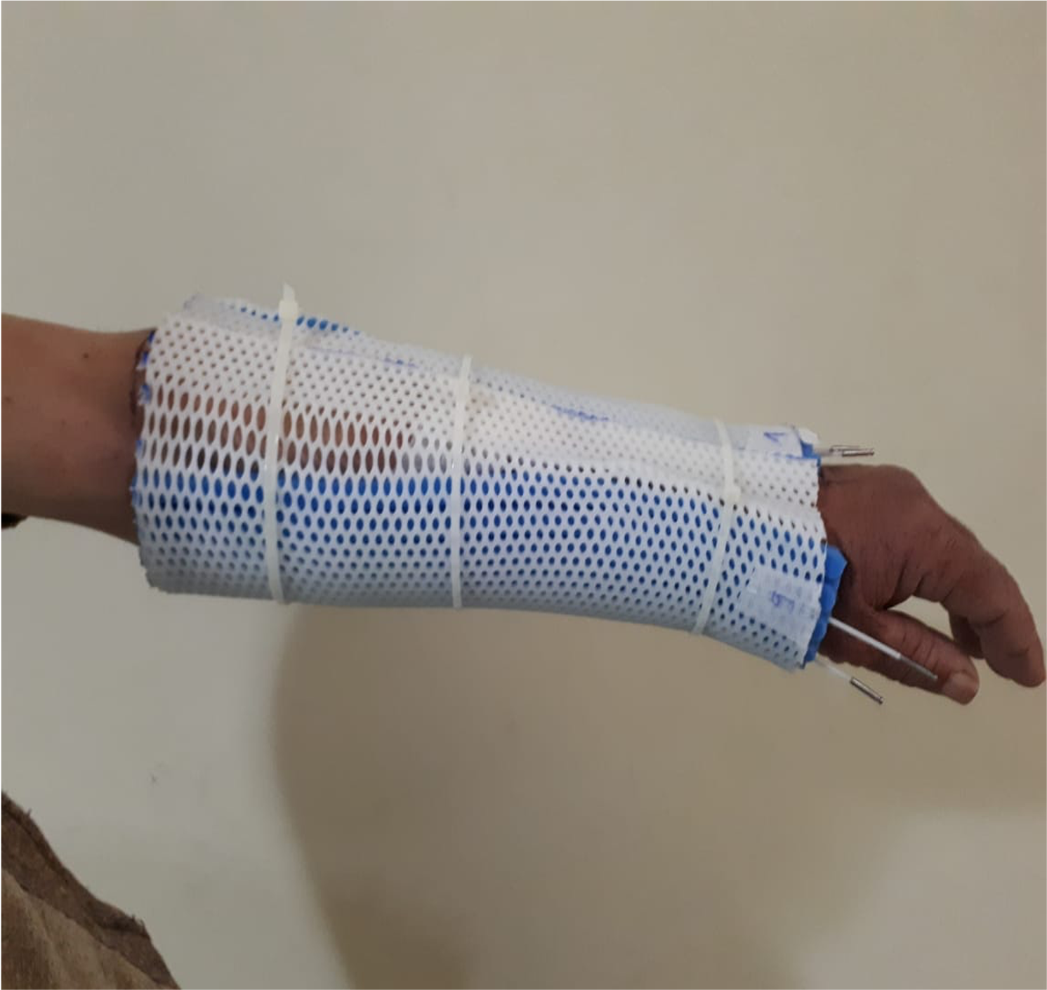
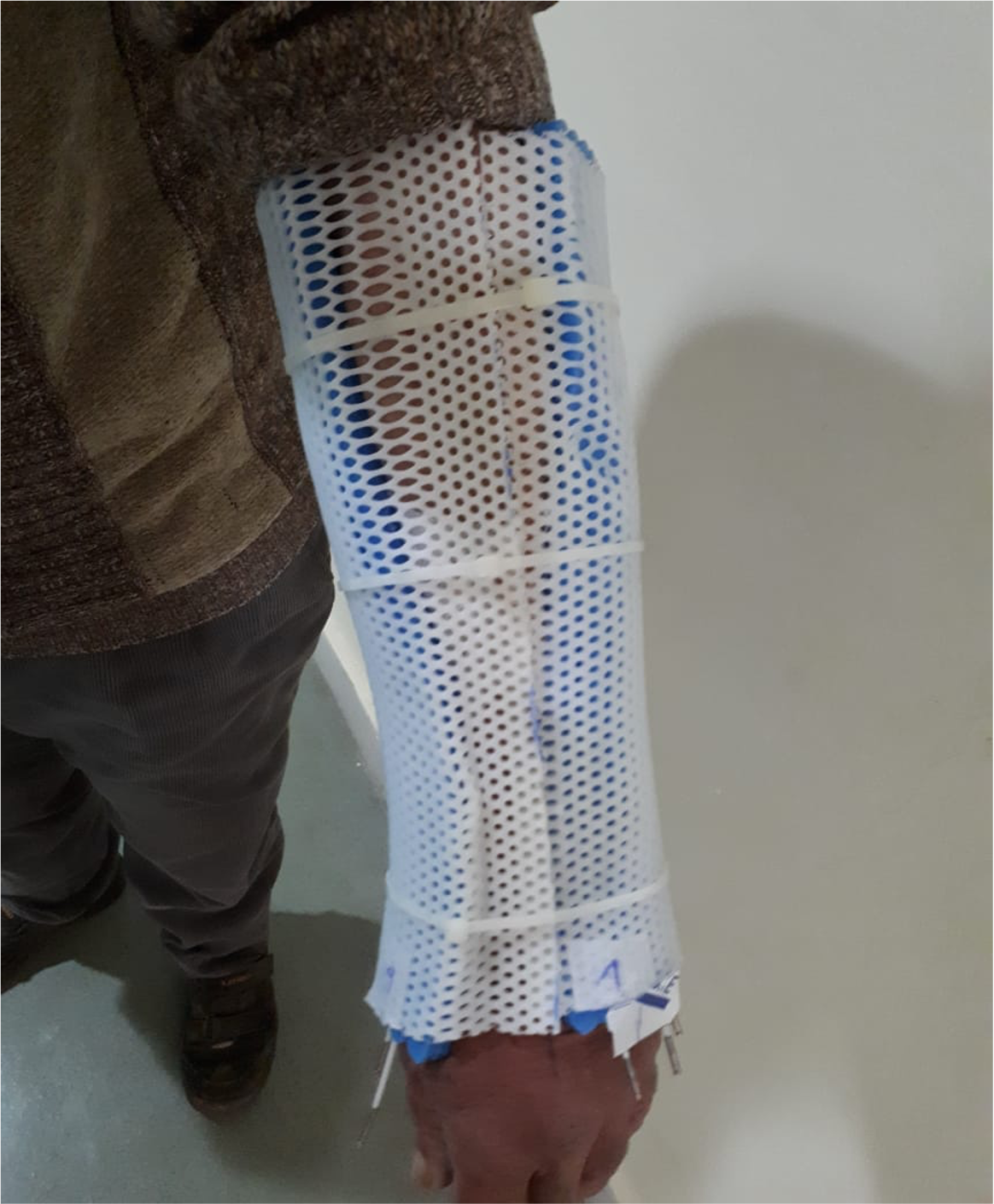
This is a retrospective study of five cases treated at the radiation oncology department of the University Hospital of FES-MOROCCO, during 4 years (2017-2021). All of our patients had a pathology examination to confirm the diagnosis and a primary treatment. RT was delivered with HDR SS-BT technique with Iridium 192. The application was made by a Catheter Flap Set type applicator by the GammaMed plus machine from VARIAN(figure 1&2). The catheter flap set is designed for treating superficial cancers, and to create a defined space between the source and the tissue, and between the source channels. It’s a highly flexible materials and easily adapted to complex anatomical shapes. The catheter channels are 2 mm in diameter, positioned on the midline of the flap with a 4 mm bolus. The catheter flap is often attached to a thermoplastic mask, to maintain a reproducible position. As for the target volumes; the GTV was the CTV and the PTV was generated from a 0.5cm margin around the CTV respecting the anatomical barriers. The dose prescription varied between 2 schemes depending on the histological type of the primary: 5x6Gy or 3x5Gy, and was prescribed at 3 mm depth. The skin surface dose was constrained to receive a maximum of 135% of the prescribed dose. V90 of the PTV had to be >85%.
Results
In our study series; we had a variety of pathology treated by this technique and the indication of RT was decided after discussing each case in MDT board: three patients with cutaneous T lymphoma (CTL); classified as T3N0M0 B0, stage IIb; as mycosis fungoid type (MF); received several treatments with no significant response. Brachytherapy was indicated in one case of desmoid tumour given the recurrent nature of the tumour and its superficial location. As for fifth case he presented a keloid scar with numerous recurrences following several treatments involving surgery, laser and bleomycin infiltration. All our patients received treatment according to plan with a daily fraction according to 2 schemes depending on the histological type of the primary. Two of our patients with CTL presented grade III radiodermatitis 15 days after SS-BT, treated with local wound management. With an average 2 years follow-up, all our patients are still alive, with no evidence of disease or recurrence.
Conclusion
We can conclude from our study that SS-BT allows to safely deposit a significantly higher dose within a tumour, with better sparing of adjacent normal structures versus plans using external beam radiotherapy (EBRT) or electron therapy.