small field electron beams: measurement based validation of the Monte Carlo calculation algorithm.
Geert Pittomvils,
Belgium
PO-1788
Abstract
small field electron beams: measurement based validation of the Monte Carlo calculation algorithm.
Authors: geert pittomvils1, erik traneus2, frederik vanhoutte1
1Ghent University Hospital, Radiotherapy-Oncology, Gent, Belgium; 2Raysearch Laboratories AB, Research & Development, Stockholm, Sweden
Show Affiliations
Hide Affiliations
Purpose or Objective
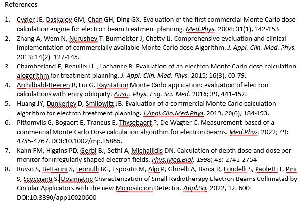
The validation of Monte Carlo (MC) based calculation algorithms for standard electron beams (E-beam) has been reported using different methods (1-6). For small field E-beams, where no lateral electron equilibrium (LSE) is obtained (7), dosimetric characterization is reported using dedicated detectors (8). We introduced small field E-beams using a similar validation process. In parallel, the obtained data is used to validate the MC Beam model for those fields.
Material and Methods
Our Elekta Synergy linacs are equipped with 5 energies and 4 applicators (6). Literature states (7) that LSE is lost for field s smaller than 6x6cm². If LSE is not achieved the R85 value will shift towards the surface and this shift increases with energy. Five symmetrical cut-outs (6x6; 4x4; 2.5x2.5; 2x2; 2x1.5cm²) for the 6x6cm² applicator were evaluated. PDD curves in a MP3 water tank were measured at a source skin distance (SSD) of 95 cm using an electron diode detector (60012), a diamond detector (60003), and a liquid filled ion chamber (31018) (PTW, Germany). Normalisation was performed by adjusting the profile using the same chamber in reference conditions. Lieteratur reported an underestimation of the virtual source distance (VSD) for small fields at extended SSD (6). Therefore, PDD data at an SSD of 115 cm were measured using the diode and liquid ion detector and compared with the RayStation 9B (RS) data (RaySearch Laboratories, Sweden).
Results
Measured PDD curves are in good mutual agreement (1D gamma; 3%, 2 mm). As expected, the R85 value shift towards the surface was observed (17 mm-12MeV; 12mm-10MeV; 8mm-8MeV; 4.4mm-6MeV; 1.3mm-4MeV). For the liquid ion detector, the response in the build-up region for small inserts is significantly lower (up to 5%). RS is able to predict the dose within the NCS-15 tolerances for the dose region at dmax or deeper but for some energies an overdosage (5%) is observed in the build-up zone. The R85 value is smaller (∑=0.82 mm) and all individual data remain within 3mm. At SSD=115 cm the measured data are in good agreement for the entire profile (3%;2mm). RS data match well around the R50 values but an overdosage is observed around dmax. Absolute dose agreement is for most fields within 3% of dose max, with maximum of 5%, but the local relative difference ranges from 1.5% to 16%.
Conclusion
This study confirms that reproducible data can be obtained but a validation using different detectors is mandatory before RS validation is started. Using the same tolerance (5%) for small photon beams, the RS MC code is validated for fields with no significant air gaps between the applicator and patient. An underestimation of the LSE in the MC model could explain the observed differences around dmax and R85 and overestimation of the VSD results in the errors observed at extended SSD. Detector based measurements are used to validate the E-MC code and help in adjusting parameters to optimize the beam model. Clinical use of small inserts is feasible with MC based treatment planning.
