Treatment of lower limb Kaposi sarcoma (KS) with radiotherapy (RT) and 3D printed custom bolus
PO-1555
Abstract
Treatment of lower limb Kaposi sarcoma (KS) with radiotherapy (RT) and 3D printed custom bolus
Authors: Kätlin Tiigi1, Rena Tiigi1, Andrei Tšižik1, Nikolai Saveljev1, Martin Tigasson1
1North Estonia Medical Centre, Radiotherapy, Tallinn, Estonia
Show Affiliations
Hide Affiliations
Purpose or Objective
To introduce lower limb KS RT with personalized 3D printed boots acting as a bolus to achieve prescribed skin dose.
Material and Methods
KS is an angio-proliferative malignant neoplasm of lymphatic endothelial origin which was described by Moritz Kaposi in 1872. It causes lesions to grow in the skin, which can occur singly, multiply in a limited area, or may be widespread. There are 4 different types (epidemic or AIDS-associated, classic, endemic and latrogenic or transplant-related) of KS defined by the different populations it develops in, but the changes within the KS cells are very similar.
Classic KS has a male:female ratio 17:1 and occurs primarily in patients >50 years old of Eastern European and Mediterranean descent. Incidence in Estonia is 0,5:100 000, it´s rare - in the last 10 years there has been 10 cases in North Estonia Medical Centre (NEMC), who treats 2/3 of all RT patients in Estonia.
For radical treatment, total dose 30 Gy is used (3 Gy x 10). Previously electron beams were used with soft gel bolus to increase skin dose and overcome the skin-sparing effect, but only lesions occurring singly were possible to treat and complicated areas, like toes, were impossible to treat accurately.
NEMC has 3D printing possibility and started to use 3D printed boots for lower limb KS RT (image 1). The CT scan was obtained with slice thickness 1.25 mm and segmented with 3D Slicer (www.slicer.org) to get the 3D model of the soft tissue. Using Blender (www.blender.org) the 3D models of the boluses were created. The boluses were 3D printed from PLA filament with 100% infill using an Ultimaker S5 3D printer.
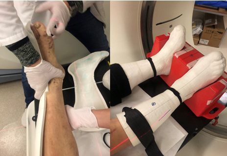
The thickness of printed bolus was 1 cm. To avoid air gaps between bolus and skin Ultrasound Gel was used. PTV was created by adding a 5 mm margin around the skin. 6 MV photon beam VMAT plans were created with the RayStation planning system to achieve 95% dose coverage to 95% of the volume. Dose calculations were made using a collapse cone algorithm with a grid 2 mm. Varian 2100EX linac with 120 MLC was used to provide the treatment.
Results
2 patients with multiple KS lesions on lower limbs have received RT with 3D printed boots and for both, treatment response is complete (image 2). The positioning and setup take more time on the linac than standard fraction slot but is easily reproducible. 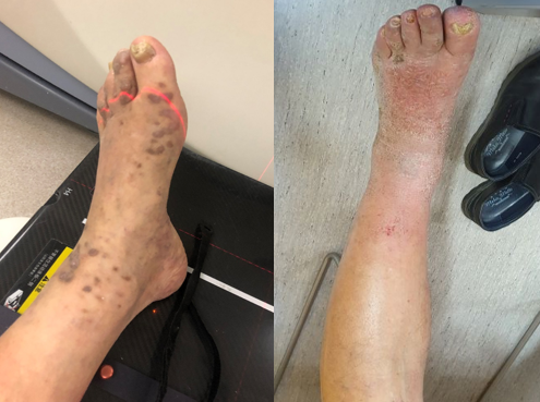
Conclusion
3D printed custom bolus is a good solution to achieve desired dose distribution in complicated regions and shows good results in terms of treatment response. In addition to classical RT equipment, a 3D printer with suitable printing material is needed, but it´s cost effective and affordable as 3D printing can be used in various disciplines in the hospital.