Automatic applicator-based image registration workflow for image-guided adaptive brachytherapy
David Neugebauer,
Germany
MO-0302
Abstract
Automatic applicator-based image registration workflow for image-guided adaptive brachytherapy
Authors: David Neugebauer1, Stefan Ecker2, Christian Kirisits2, Nicole Eder-Nesvacil2, Petra Trnkova2, Dietmar Georg2
1Heidelberg University Hospital, RadioOnkologie und Strahlentherapie, Heidelberg, Germany; 2Medical University of Vienna, Department of Radiation Oncology, Comprehensive Cancer Center, Vienna, Austria
Show Affiliations
Hide Affiliations
Purpose or Objective
Image-guided adaptive brachytherapy (IGABT) is a crucial part of the treatment of patients with locally advanced cervical cancer, following radiochemotherapy.
In many clinical settings, the treatment is applied with two applicator insertions, each serving two fractions of high-dose-rate BT. Subsequent fractions are monitored by three-dimensional imaging to assess interfraction motion of organs at risk (OAR). Significant implant shifts or large anatomical variations lead to new treatment planning, otherwise, the original treatment plan is reapplied. Optimally, the effect of anatomical variations is quantified in terms of a dosimetric difference, functioning as a basis for decision-making. However, with existing methods this is a time-consuming process which limits its clinical feasibility.
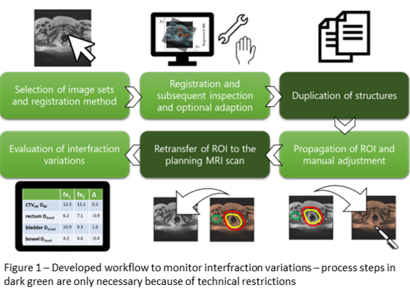
In this study we establish a semi-automatic workflow to monitor interfraction variations of OAR using the scripting interface of a commercial treatment planning system (TPS) (RayStation, RaySearch Laboratories, Stockholm, Sweden), and investigate the suitability of different rigid image registration algorithms.
Material and Methods
10 patients were treated with magnetic resonance (MR) IGABT. Clinical para-transversal MR-series (slice thickness 5mm, in plane resolution 1.17mm) and treatment plans (Oncentra Brachy, Elekta, Veenendaal, The Netherlands), including reconstructed applicator positions for two fractions were available for all patients, with the same applicator in-situ.
Applicator contours, which are usually not available were generated with an Elekta Applicator Slicer research plugin and exported as standard DICOM structures.
A semi-automatic workflow script for interfraction variation assessment, was developed using Raystation 12A.
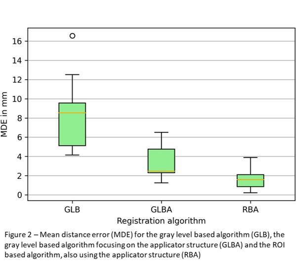
In order to identify the optimal workflow for automatic rigid BT image registration, the following registrations algorithms, available in the TPS, were investigated: the general gray level based algorithm (GLB), the gray level based algorithm focusing on the applicator structure (GLBA) and the ROI based algorithm (RBA), also using the applicator structure.
Results
The established workflow to semi-automatically quantify interfraction variations in BT, subject to some technical limitations in the TPS, is schematically depicted in Fig. 1.

Results of the comparison of different registration algorithms are summarized in Fig. 2. Evidently, the presence of the applicator structure could significantly boost registration accuracy for the RBA algorithm (MDE 1.59 mm), in comparison with GLB (MDE 8.40 mm) and GLBA (MDE 3.39 mm). 
Conclusion
The developed workflow represents a promising proof of concept for fast quantitative evaluation of interfraction variations, the largest source of uncertainties in dose reporting in HDR-BT for cervical cancer. Our data supports the assumption that for future clinical application of such an automated workflow, the automatic prediction of the applicator structure on MR images, e.g. by a neural network, would be required.