Course report
Imaging for Physicists - PDF Version
14 April – 19 May 2021 - Online
Course director:
Rob Tijssen, physicist, Catharina Hospital Eindhoven, The Netherlands
Could you please briefly introduce yourself?
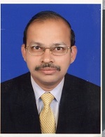
My name is Sivakumar Somangili and I have worked in the field of medical physics for the past 20+ years. I am a radiation oncology medical physicist accredited by the Australian College of Physics in Science, Engineering and Medicine (ACPSEM). I currently work at the Sir Charles Gairdner Hospital as a senior medical physicist in Perth, Western Australia. As a radiation oncology medical physicist in one of the busiest public hospitals in Australia, I am expected to be involved in multi-tasking; I have to attend to my regular clinical duties as well as to discharge my obligations in short/long-term projects and be involved in training medical physics registrars. My career has taken me from India to Oman and then to Australia, where I have been since 2009. I have had learning opportunities such as commissioning Truebeam linear accelerators, the Eclipse planning system, the Brainlab stereotactic system, nucletron brachytherapy, Varian portal dosimetry etc. My hobbies embody my spirit of living in the midst of a heavy schedule. My primary hobby, almost verging on passion, is to listen to any percussion performance. My favourite outdoor preoccupation is cycling, alone or with company. I also love to watch and wonder at scenes of nature such as sunsets and free birds in the sky, and I also take an immense interest in aviation.
Why did you choose to attend this course?
I want to develop sound fundamentals in different imaging modalities, from both the teaching and clinical points of view.
What aspects of the course were the most interesting and why?
I most enjoyed the pre-recorded audio lectures. The quality of the recordings was good, while the topics chosen and the contents were well balanced. Listening to these lectures (especially on MRI and nuclear medicine) helped me to be able more fully to answer any questions that our trainee physics registrars might pose. In particular, the contribution by one of the presenters of different MR images of fruit was a perfect blend of novelty and the subject.
Did the course activities improve your knowledge and skills in the relevant subject?
Certainly I would say yes but primarily in terms of the theory. I hope now I am in a position to identify artefacts in MRI and correlate them with the likely cause.
Did the course meet your expectations? If so, how?
The course contents were well balanced in terms of basic theory of different imaging modalities, their limitations and the shortfalls/pitfalls that are associated with each one in different clinical scenarios. The follow-up Q&A sessions and quizzes were a well-thought-out initiative by the organisers to ensure that the participants gained knowledge and understood the clinical significance of all imaging modalities.
List three important ‘takeaways’ following the course.
- Position emission tomography (PET) is reliable in nodal staging in lung cancer.
- Do not rely exclusively on deformable image registration in PET computer tomography (CT).
- There are pros & cons in experimental and stoichiometric Hounsfield unit CT calibration.
How will what you have learnt be implemented in your daily job/ clinical practice?
Based on the knowledge gained, I am working out how to identify the shortfalls in our current MRI and PET quality assurance programme pertinent to radiotherapy and how to implement changes through collaborative work with our radiology department.
How would you encourage someone who has never been to a course run by the European SocieTy for Radiotherapy and Oncology (ESTRO) to join this course next year/ in two years?
Based on my experience in the course, it would not be difficult for me to convince the other person with relevant qualifications that the course would update their knowledge and enhance their confidence, as has happened in my case.
I see this course as an additional learning opportunity to build a strong knowledge base in all imaging modalities that are used in radiotherapy, to meet experts and to develop purposeful, professional networking opportunities. I would recommend this course to medical physics registrars and physicists who are interested in understanding the significance of imaging in current radiotherapy practice.

Sivakumar Somangili
Senior medical physicist/ stereotactic radiosurgery
Sir Charles Gairdner Hospital
Perth, Western Australia
Sivakumar.Somangili@health.wa.gov.au