3D printing of individual skin brachytherapy applicator: design, manufacturing, and early clinical results
Bieleda G, Chichel A, Boehlke M, Zwierzchowski G, Chyrek A, Burchardt W, Stefaniak P, Wisniewska N, Czereba K and Malicki J
Contemp Brachytherapy 14 205-14
What was your motivation for initiating this study?
A common problem in superficial brachytherapy of the head and neck region is the lack of a well-fitting applicator. Either the applicator does not adhere tightly to the lesion's surface, or else it is impossible to achieve a dose distribution that is acceptable in the target volume and the critical organs simultaneously. Most often, this is a problem comprising both elements. A solution often used in brachytherapy is to prepare individual moulds. However, this requires a well-equipped mould room and highly skilled, manually proficient personnel, and the result is highly dependent on the unique characteristics of the technician. We decided to develop a technique for producing individual applicators using 3D printing technology to circumvent these obstacles. We wanted to make the preparation of the applicator as intuitive as possible and not require the user to have programming skills. It was also important that the applicator could be designed in the treatment planning system and verify the result obtained without unnecessary transfer of data between different programmes.
What were the main challenges during the work?
The main technical difficulties consisted of extracting information from the treatment planning system about the geometrical shape of a putative applicator. Another obstacle was determining the optimal catheter path for the radioactive source and placing the two structures in the same coordinate system. It was also necessary to subtract the two structures from each other to create channels of the correct shape and diameter in the applicator so that commercially available catheters could be quickly but firmly inserted. Verifying the dose distribution around such a fabricated applicator was also a non-trivial issue, but this had been solved in our previous studies.
What are the most important findings of your study?
The most important thing we were able to prove is that it is possible to make individual applicators for contact brachytherapy in anatomically challenging locations. This approach does not require a significant financial investment, hiring specialised personnel, or dedicating additional space for this purpose. The entire process of designing the applicator is carried out in the treatment planning system, and only the finished shape is exported to free external software that works securely without an Internet connection. Printing of the applicator is possible on any 3D printer. It does not matter what material the printer works on, as only the physical parameters of the material used need to be verified. In addition, we prepared an instructional video on how to prepare such an applicator in your brachytherapy department. Such a supplement to the article is available on the publisher's website. Last and equally important, the proposed and tested workflow enables the doctor and physicist to invent, design and prepare the most convenient treatment plan before the final applicator is manufactured and put on the treated lesion. This approach was not possible in our best previous practice.
What are the implications of this research?
It has become possible to prepare clinically and dosimetrically acceptable treatment plans for skin cancer patients for whom the alternative could be a mutilating

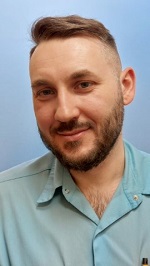
Grzegorz Bieleda, PhD, medical physicist
Electroradiology Department,
Poznan University of Medical Sciences,
Poznan, Poland
and
Medical Physics Department,
Greater Poland Cancer Centre, Poznan,
Poland